But 1963 may not be the earliest date in which a cell exhibiting stem cell characteristics is mentioned. The term is mentioned in a 1932 medical journal, but the reference to what we know as stem cells today seems inappropriate. And then there are older references to stem cells back to 1905.
One thing is clear, however, we had little understanding of what these cells were and their potential.
Today we know about stem cells. The Mayo Clinic’s website calls them the “body’s raw materials,” cells from which all others can be generated. But what makes them so different from all those other cells in our bodies? Our bodies contain 200 known varieties of cells. Each, with the exception of blood cells, has a nucleus, mitochondria plus other organelles, a common chemical soup, and consistent structural features.
Yet there is no such thing as a typical cell. All cells pretty much do the same thing. They divide until their ability to do it stops. At that point, they die. We lose 96 million cells per minute but we also gain that same number in the same amount of time. The behaviour of each cell is determined by instructions given by genes located on chromosomes within the cell nucleus.
When we humans start out, our first cell is a fertilized egg combining DNA from both of our parents. The egg divides to eventually form a ball of cells we call an embryo. And the modern story of stem cells begins with what scientists first discovered about embryos. They contained cells that were non-specific but capable of being instructed to differentiate to turn into us, producing our bone, muscle, organs, brain, nerves skin, and nails. With all that differentiation leading to the 200 known varieties of cells we find in our bodies today, scientists overlooked one. Stem cells didn’t cease to exist when the embryo began to differentiate. They very much exist within all those differentiated organs and tissues that form us. You can find them in muscle, bone, organs, and fat. In fact, body fat may prove to be the best source of harvestable stem cells that we can use to cure so many of the chronic conditions and diseases we humans encounter.
Most of us when we think of stem cells, we think embryonic. That’s because that’s where the science of stem cells first started. To make embryonic stem cells researchers use a technique called somatic cell nuclear transfer. It involves manipulating donated eggs by removing the original DNA contained in the nucleus and replacing it with DNA from a donor adult cell. Cell division then follows until an embryo forms. The embryonic cells, about 100 of them on maturity, are then harvested and the embryo destroyed.
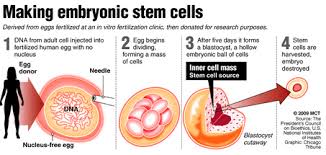
It is this use of human embryos that has created the controversy associated with stem cell research. For those who aspire to the belief that life begins at conception, harvesting and destroying an embryo is seen as murder. Lobbying by religious groups soon led to restrictions in the science in the United States with only a limited number of embryonic stem cell lines allowed.
But the promise of these cells and their ability to differentiate into any type of tissue has kept research laboratories busy in places other than the United States where more than 300 existing lines of human embryonic stem cells being cultivated in Canada, Sweden, France, Spain, and the United Kingdom. These lab-grown embryonic stem cells represent one branch of stem cell research. And because the cell populations are artificially cultured in laboratories there are some significant problems that can happen.
Consider evolution, the engine that drives speciation. From reptiles came dinosaurs, mammals, and birds. How did that happen? Mutation. And mutation worked best when there were large populations of individual species who through breeding, living and dying further differentiated and survived.
Now apply that same observed principle to laboratory populations of continuously dividing limited lines of stem cells. Just like in an isolated population of animals with a limited amount of DNA to share, mutations can produce undesirable results. In the case of laboratory-grown stem cells, a cell meant to cure could because of undesirable changes kill.
In addition, growing a large enough population of embryonic stem cells to cure people is an expensive proposition. The science, therefore, needs to find a better way to use the potential regenerative capacity of these cells.
Finding a new source of stem cells is critical. And lo and behold they are there, in our bodies, lurking among all those differentiated cells which came after conception.
In Part 2 we’ll continue the story, describing how readily available stem cells existing within us represent a medical revolution in tackling a wide variety of diseasee challenges from autoimmune to cardiac, hepatic, pancreatic, gastrointestinal, respiratory, neurodegenerative, ophthalmologic, spinal cord, skin, osteo, and more.
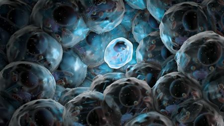
Many diseases we humans experience are caused by malfunctioning cells. Stem cell research focuses on replacing those malfunctioning cells with healthy ones conceived from the patients themselves. In this case somatic cells, those derived from the skin of two adult male donors, were injected using a process known as somatic cell nuclear transfer (SCNT). The result, the generation of donor-specific pluripotent stem cells that can be differentiated into different tissue types for use in treating a number of diseases.
Stem cells derived from embryos have shown promise in treating eye conditions like macular degeneration and dystrophy. But now cells derived through SCNT could conceivably be used to treat Alzheimer’s, Parkinson’s, Type 2 Diabetes, Osteoarthritis, or immune system diseases.
I suffer from osteoarthritis and am a candidate for knee replacement. It is conceivable that I could donate skin cells which could then be turned into cartilage and injected into my knee joint to restore what I have lost over the years. Currently clinical trials by Advanced Cell Technology focus on treating macular degeneration and dystrophy. Well I’ve held off getting the knee replaced, so maybe I’ll wait a bit longer and see if there will be a clinical trial for my condition in the near future.
SCNT can also restore damaged heart muscle in patients who have suffered a heart attack, or can replace the skin of burn victims. In fact the technology has the potential to change how medicine treats acute trauma cases.
The research appeared in the April 17 edition of the journal, Cell Stem Cell.

Visit the Navigant site to subscribe and receive more of their insightful reports and predictions about the future.
But today I am keeping my headlines focused on recent biomedical announcements that I hope you find interesting as well. They are:
Stem-Cell Therapy and Non-Lethal HIV Injection Produce Two Very Different Cures
Two HIV stories were in the news this week. The first is a sequel to one I wrote about some time ago involving a gentleman named Timothy Brown, who may be the first person to be “cured” of HIV after receiving a bone marrow transplant in 2007. Well now stem cell transplants to treat lymphoma may have “cured” two more HIV patients. During the transplant both patients remained on HIV medications but eight months later both are no longer on their meds and are doing well according to Timothy Henrich, Harvard Medical and Brigham and Women’s Hospital in Boston. The HIV virus may still be around but it is not showing up in any blood work and that’s a pretty good sign.
The second story concerns a 7-year old suffering from acute lymphoblastic leukemia since age 5. After multiple rounds of chemotherapy, little Emily Whitehead, was no better. But her parents found out about a gene therapy regimen being tried at Children’s Hospital of Philadelphia and took her there. The treatment is called T-cell immunotherapy and involves delivering new instructions to these cells which play a major role in fighting disease. Because viruses can lock on to receptors and penetrate cellular membranes they can be used to deliver instructions to a cell. In this case the transmitter chosen was a disabled AIDS virus containing new genetic material to switch on the T-cells capability to identify leukemia cells. The T-cells once infused with these genetic instructions get transfused back into the patient where they begin attacking the leukemia. Emily is one of 10 children who have received T-cell immunotherapy. She is doing well as are many of the others who have benefited from an AIDS virus repurposed for good.

Vitamin D Plays a Role in Surgical Recovery
I take several thousand units of Vitamin D every day because I live in a northern country where half the year there is less than adequate sunlight to restore my Vitamin D reserves. It along with calcium supplements helps me maintain my bones.
But we are finding out that Vitamin D which we mostly get from sunlight and not supplements has another purpose. A study done at Children’s Hospital of Eastern Ontario in Ottawa, Canada headed up by Dr. Dayre McNally looked at 58 children undergoing cardiac surgery to correct congenital heart defects and found that almost all saw levels of Vitamin D drop post-operatively. That drop correlated to longer recovery periods.
The researchers concluded that the drop in Vitamin D was associated with usage of cardiopulmonary bypass machines during surgery, previously an unknown side affect. As a result of their discovery the hospital is developing a new protocol to optimize Vitamin D in children prior to and post-operatively to maintain high levels. This, they hope, will mean reduced recovery times and faster healing. These findings have ramifications well beyond heart surgery as researchers are finding the vitamin also plays an important role in the health of many other body parts including lungs and our immune system.

New Exoskeleton Concept Replaces Casts to Help Heal Fractures
When I was 20 I broke both of my legs. I was in and out of hospital and plaster casts for the better part of a year-and-a-half as my bones healed. From personal experience I can tell you just how uncomfortable and unhygienic plaster casts are. So when I was told about CORTEX I just had to find out more.
What is it? CORTEX is an exoskeletal cast that is fully ventilated, light, lets the patient wash the recovering limb, and is made of fully recyclable materials. It is the product of Jake Evill, an industrial design graduate from Victoria University in New Zealand. It resembles a honeycombed exoskeleton and is individually created using X-rays, a 3D scan of the fractured limb, and a 3D printer to generate the custom cast including snaps and fasteners for application and removal. The steps are illustrated below.
Mr. Evill is looking for investors to help him bring CORTEX to market. If you are interested visit his web site.

3D Printing of Stem Cells Helps Build Bones
More 3D printing in the headlines this week. In London, in the United Kingdom, this week, a new technology is on display at the Summer Science Exhibition. It’s a 3D bioprinter that is using adult human stem cells to create bones. The printer’s ink combines a polymer, polyactic acid, and an alginate to create an object that is equivalent in shape and size to a bone. The object is coated with human stem cells and becomes a scaffold for those cells to use to regenerate bone. When implanted as the bone regenerates the scaffolding degrades disappearing within a few months. What’s left is perfect replacement. On display at the Exhibition this week is a lower jaw and skull made using the 3D bioprinter. The latter eerily resembles the crystal skull of Indiana Jones lore (see below).

Tiny Human Livers Fabricated Using Stem Cells
For more than a decade it seems stem cell research has crept forward as U.S. restrictions on stem cell lines held up progress. But it seems from the headlines of the past week that the dam has burst and we are seeing great strides in the field. And one of these breakthroughs comes from Japan where researchers are using human stem cells to create human livers.
As reported in Nature this week, human induced pluripotent stem calls (converted skin cells) have been successfully used to create functional working livers, albeit very tiny ones. The researchers describe their work as proof-of-concept for developing organ-bud transplantation in the burgeoning field of regenerative medicine.
Imagine taking human skin cells from someone suffering from liver failure, inducing those cells to revert to a stem cell state and then stimulating them to turn into healthy liver cells. That’s exactly what the researchers are doing. These little liver buds (see image below) work just like your liver, capable of filtering body fluids and metabolizing drugs. Researchers believe they can scale the technology enough to regenerate as much as 30% of a patient’s liver. Talk about life extending technology!

A Postscript
This week we focused our headlines on biomedical stories. Who knows what the 21st century will bring us next week? But I’m glad you could drop by and spend the time here and look forward to your comments and questions.
– Len Rosen
]]>
Adult Stem Cell Research – Going back to 2002 NASA started experimenting with human stem cells in weightlessness creating a “rotating wall bio-reactor.” Today that technology has been licensed by Regenetech to continue the work. Regenetech acquired exclusive rights to 11 NASA patents and is using the bio-reactor which it calls the Intrifuge System. How does the bio-reactor work? In the simulated weightless, microgravity environment cells form large clusters that resemble the way tissue normally grows in the body. The constant rotation gently mixes cells and nutrients without damaging cell growth and interaction. Currently the system in cooperation with medical institutions is assisting in the study of human colon, prostate, breast and ovarian tumors. The bio-reactor is also being used for experimentation in the building of cartilage, bone marrow, heart muscle, skeletal muscle, pancreatic islet cells, and liver and kidney cells from adult stem cells.

Digital Fly-by-Wire – or as it is known by acronym, DFBW, was first developed by NASA engineers back in 1972. The aircraft, a modified F-8 Crusader was fitted with with a digital command console (seen below) and wires to replace hydraulic controls. The wire controllers were less vulnerable to damage under the stresses of battle conditions or space flight and much lighter in weight. The computer-aided guidance meant the pilot could rely on the controller to make decisions to achieve stability and a smoother ride. DFBW was available on Apollo and became standard on the U.S. shuttle. Today DFBW systems are a feature of almost all commercial aircraft.

UVA Filters – The commercial eye industry owes a lot to NASA for its development of lenses and lens coatings that absorb 99% of hazardous light including UVA and UVB. For NASA astronauts light filtering in the harsh environment of space was an absolute necessity. Short-term eye exposure to UV radiation causes photokeratitis, an inflammation of the cornea. Long-term exposure leads to cataracts, retinal damage, and eye lesions. Today contact lenses, regular eye glasses and sun glasses include UVA and UVB protection.


Aerodynamics and Fluid Dynamics – Today’s trucks and automobiles owe a lot to NASA’s engineers at the Dryden Flight Research center. Before NASA only racing car designers talked about drag coefficients and aerodynamics. But when NASA started developing rockets and spacecraft the ideas they incorporated into design soon found a new audience dealing with rising costs of transportation fuel. Hence we see today faring and air dams on semi-trailer trucks that save hundreds of liters of fuel over a single trip, rounded shapes, grill-closing technology to reduce drag, and other design details to reduce turbulence and drag coefficients.

Food Supplements – From Tang to micro algae, NASA has done more than most in developing new foods and food supplements to help it achieve the goal of keeping astronauts alive in space. In Elena Malykhina’s article she specifically talks about Martek Biosciences, who has through licensing of patents and scientific breakthroughs from NASA’s Closed Environment Life Support System, introduced a range of nutritional products under the brand life’s DHA. Developing strains of micro algae that produce oil rich acids and nutrients, life DHA products include supplements for infant, child, maternal and adult health. DHA is now found in nationally distributed foods made by companies such as Kellogg and Yoplait. Look at the picture below to get a good sampling of NASA-inspired food products.

So there you have it, the contributions that NASA has made through its space research to those of us who may never go into space but certainly are beneficiaries of what the program has produced.
]]>What the Heck is the Higgs Boson and What Does it Mean for Us in the 21st Century?
When scientists get excited about a discovery like the one announced this week it is hard not to jump on the bandwagon and acknowledge the progress made to unlock the mysteries of the Big Bang, matter, antimatter, dark matter, energy, dark energy and the quantum sub-atomic realm.
But what does the Higgs Boson particle discovery mean practically speaking? To answer this question you have to appreciate what this particle means to other sub-atomic particles. The Higgs Boson is the attractor that makes sub-atomic particles associate with each other. It acts like an invisible force field giving other particles their mass. Without the Higgs Boson electrons wouldn’t exist, and without electrons atoms wouldn’t exist, and without atoms we wouldn’t exist.
Consider the importance of electrons to our technical society (all of our energy and power generation is based on what electrons do) then it’s good to know our understanding of how things work in the universe is confirmed by this discovery.
Billions of dollars have been spent searching for the Higgs Boson as nations cooperated to build the Large Hadron Collider. And now with it built the Collider can make important contributions to science, and feed technology innovation in a number of fields from biomedicine, to manufacturing, to energy, computing and space science.
It’s hard not to argue that advances we have made in scientific discovery in the past have always led to new technology. So hats off to the scientists at CERN and to future discoveries from the Large Hadron Collider. And before I end this topic I leave you with the following:

Biomedicine Update: Move Over Higgs Boson as Researchers Discover Holy Grail of Tissue Engineering
Newts and other amphibians can regenerate lost limbs. Some reptiles can regenerate lost tails. Even humans can regenerate bone to heal fractures. But regenerating lost brain cells, heart muscle, or insulin-producing pancreatic cells has been only a dream until now.
Well according to the Journal, Comptes Rendus Biologies, scientists at the University of Paris Descartes and University of Maryland, adult bone marrow stem cells harvested from mice were used to generate multiple tissue types for use in cell replacement therapy.
If the mouse study can be replicated in humans we will have the ability to replace damaged organs without transplants and the need for immune suppressants.

Energy Update: Paint on Batteries
What if you could spray paint batteries on to any surface from a ceramic tile to a piece of paper? That’s exactly what researchers at Rice University, in Texas, hope to achieve. They have developed technology to spray-paint battery structures on surfaces. The anode and cathode, that is the negative and positive halves of the battery are kept apart using poly-methyl methacrylate. A sequence of five separate paint layers, each no more than 0.5 millimeters in thickness, makes a battery with efficiencies equivalent to the traditional lithium-ion batteries we use today in portable electronics. The flexibility these batteries provide should make them attractive to consumer-electronics and industrial manufacturers.

Materials Science Update: New Adhesives Created from Barnacles
Current adhesives are typically made from petroleum byproducts. But nature demonstrates adhesives with remarkable bonding capability, almost impossible to break. The Buoy Barnacle, Dosima Fascicularis, produces an adhesive that allows it to stick almost anywhere in the marine environment. Naturally its adhesive can cure under water.
So a research team at the Fraunhofer Institute for Environmental, Safety and Energy Technology in Bremen, Germany, is studying the barnacle to isolate the amino acids and proteins that constitute the properties of the adhesive with the goal to use it in medical applications such as closing surgical incisions, and supporting pins and screws in repairing bone fractures.

Space Science Update: Could We Build the U.S.S. Enterprise Today?
An engineer has proposed that much of the technology needed to build a starship like Enterprise exists today not including warp engine technology and transporters. For example we have technology today that was seen as futuristic in the 1960s when Star Trek first appeared on television. Touch screens, communicators, sliding doors, talking computers, and replicators (think 3D printers) are available to us today. Even impulse engines represent technology currently under development in NASA’s Advanced Propulsion Physics Laboratory.
Unlike the TV Enterprise, the one proposed would use a rotating structure in the saucer section of the spacecraft to create artificial gravity.
You can check out the website at BuildtheEnterprise.org.

A Quick Primer on Heart Disease
Humans get two types of heart disease: congenital (that is born with) and acquired (disease that develops over time). Congenital tends to be structural. It can express itself in abnormal anatomy or physiology. Acquired begins with normal anatomy but physiology changes as the heart becomes diseased. Both can lead to heart failure which you can live with for a very long time or heart attacks which can lead to death.
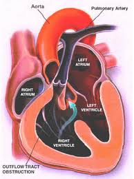
Structural defects to the heart and blood vessels in congenital heart disease can be repaired surgically, missing valves implanted and blood flow restructured using human grafts and artificial materials. Drugs can help regulate heart rhythm or improve the pumping efficiency. Pacemakers can help overcome structural problems that impact the electrical system that regulates heart rhythm.
Acquired, which represents the bulk of what we normally think of as heart disease, can also benefit from surgical intervention, drugs, pacemakers and other devices.
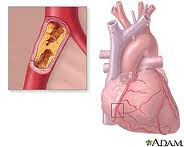
What causes heart disease? Lifestyle and inherited genes are the principal cause with 30 to 60% of the risk associated with the latter when it comes to acquired, rather than the former. Recent international studies identified 17 new genetic variants linked to increased heart disease risk. Five of these were associated with genetic regions that govern lipid metabolism processes, genetic controllers that impact the build up of fatty deposits in arteries. Ten others were in genes involved with other diseases and traits such as autoimmune diseases like Celiac and Type 1 Diabetes, cerebral and abdominal aneurysms, and lung cancer. What this means is there is no single genetic “smoking gun” that defines whether a person is likely to get heart disease in his or her lifetime. Instead we have a complex of genetic markers.
New Therapies for Repairing Bum Hearts and Blood Vessels
Stem Cells
In past blogs we have talked about stem cell therapy. Because stem cells have the potential to develop into a range of tissue types we can use them to regenerate and replace diseased tissue.
Cellular Dynamics International (CDI), is an American company located in Madison, Wisconsin, that is experimenting with the reprogramming of adult somatic cells to make them revert to a pluripotent state so that they can grow into any cell type needed for doing repairs to the body. These are induced pluripotent stem cells or iPS. Since 2008 CDI has used iPS cells to manufacture cardiomyocytes (heart muscle cells), freezing and sending them to pharmaceutical companies doing research on new drugs to treat heart disease.
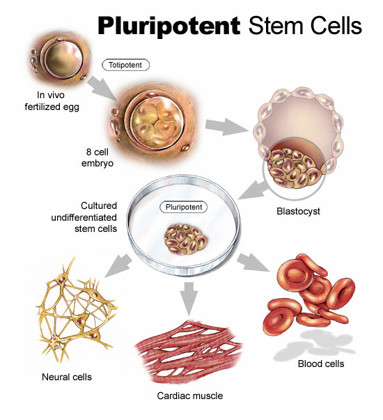
But iPS cells have a brighter future if we can work out some of the bugs. Those bugs include faint traces of chemical residue from the parent cells used to create the reverted cells. Those residues can lead to mutations and as we have already written in previous blogs, mutations can lead to runaway cancer cell proliferation.
You can create iPS cells from almost any human tissue source. If a patient with a damaged heart provides a tissue sample, the cells from the sample can be induced to revert to iPS cells. They then can be transplanted back into the patient at the location requiring treatment. iPS cells from the donor are not rejected because they are from the donor who is also the recipient. The iPS cells in their reverted state can take on the characteristics of those cells that are in proximity to them and start to divide. iPS cells in the heart can generate heart muscle. iPS cells in the spine can generate nerve cells. CDI charges pharmaceutical companies $1,500 for 1.5 million iPS cells. That’s not cheap but the potential for iPS as an alternative therapy to transplants makes this a desirable area of ongoing research.
In research being conducted in Bristol, England and Glasgow, Scotland, Doctor Costanza Emanueli, along with a team of scientists are studying a variety of approaches to treating heart attack victims including using embryonic stem cells to create blood vessel cells to inject into patients. The method of delivery is a skin patch.
Gene Therapy
Researchers are looking at ways to tweak DNA to repair injured hearts. This involves manipulating a variety of genes that impact muscle and blood vessel growth.
Dr. Emanueli’s team, mentioned in the previous section, is studying boosting levels of nerve growth factor (NGF) to improve heart attack survival rates. In studies with mice the death rate from heart attacks was reduced by half using NGF. The gene responsible for NGF was attached to a specially engineered virus and delivered into the hearts of mice. Mice that received the NGF gene showed much improved circulation and heart function. In previous studies NGF was known to regrow nerves but in this case it encouraged new blood vessels to grow inside the injured heart muscle.
In another study conducted at Harvard’s Beth Israel Deaconess Medical Center, the gene C/EPB-beta spurred the growth of cardiomyocytes. This study showed that a genetic trigger responding to physical exercise turned on a molecular pathway to get cells to start dividing. For a very long time cardiologists could not find evidence of cell division in adult heart cells. Hearts under stress or subject to exercise would swell but cell proliferation in these enlarged hearts was poorly understood.
Heart muscle enlargement, called hypertrophy, can be associated with disease or with what is referred to as “athlete’s heart.” In the study cardiomyocyte growth was attributed to molecular events rather than factors such as high blood pressure.
Reprogramming Heart Cells After a Heart Attack
Heart attacks in the past have caused irreparable damage to heart muscles. What causes them? The usual suspects are accumulations of deposits on the walls of arteries that supply the heart with blood. When an artery is partly to fully blocked oxygen and nutrients are cut off to the area next to the blockage. This kills the muscle near the blockage affecting the heart’s ability to beat. But what if the section of damaged heart muscle can regrow?
Professor Paul Riley, of Oxford University’s Department of Physiology, Anatomy and Genetics, and his team of researchers are looking at a layer of cells known as the epicardium. The epicardium is the inner wall of the sac of tissue known as the pericardium that surrounds the heart. The embryonic epicardium contains many types of cells used by the heart and circulatory system including blood vessels and heart muscle. But after birth the epicardium gets quiet and stops generating these tissues. Dr. Riley and his team looked at ways to switch the epicardium cell generation back on. In mice studies they achieved just that by administering doses of Thymosin Beta 4, a natural occurring protein, to stimulate the embryonic genes in the epicardium to create cardiomyocytes. The researchers found that administering Thymosin Beta 4 before a heart attack proved to stimulate faster regrowth of heart muscle tissue.
The team continues to hunt for other naturally occurring molecules that can stimulate heart muscle and blood vessel growth. Along with drug and interventional therapies to clear arterial blockages, administering these proteins pre-emptively could create a bank of healthy new heart muscle to speed recovery should an attack occur.

Her journey from the petri dish to birth began as a cell taken from a mammary gland of a 6-year old female donor. The technique included putting the cell into a suspended state to extract its DNA. A host egg cell came from another female donor. With the egg cell nucleus removed the DNA from the mammary cell was inserted. Then an electric current was applied to simulate the energy accompanying fertilization and embryonic development. After 148 days, Dolly was born.
It wasn’t all that simple a task. The researchers tried this with 277 fused eggs. Only 29 embryos survived and of these 13 were successfully implanted, but only one, Dolly, was born.
Cloning is not new to nature, just new to humanity. Many creatures practice asexual reproduction, or parthenogenesis, producing exact copies of themselves. Many plants reproduce themselves by sending out roots laterally and sprouting exact DNA copies of themselves. Researchers studying Aspen trees in British Columbia, however, report that this form of cloning leads to an increasing likelihood of creating some bad genetic material. We call altered genes mutations. The scientists studying Aspens counted the accumulated mutations of 20 different male Aspen trees and noted that cloned trees were less hardy than their parents. The same can be said about Dolly. She aged prematurely and passed away after 6 years.
Dolly wasn’t the first vertebrate to be cloned. That honour belongs to a carp, cloned in 1963.
The first cloned mouse, named Cumulina, was created in 1997 and died in 1999.
Mira the goat, born in 1998, was actually 3 goats, all identical. Their DNA was modified so that their milk produced recombinant human antithrombin (rhAT), a protein that prevents blood from clotting.
Cow clones, made in Japan, appeared in 1998. Recently, a newspaper in the United Kingdom described some of the challenges related to cloned farm cows with reports of pain, ill-health, organ defects and gigantism.
The first cloned guar (an Indian bison) named Noah was born in 2000 and died 48 hours after birth.
Pig clones have popped up in the United States, the United Kingdom, Japan and other locations, the first appearing in 2000. In 2001 scientists produced the first pig clones genetically modified to grow organs suitable for human transplantation. This feat was accomplished by knocking out a pig gene that produces the coating on organs containing sugar molecules that trigger acute rejection when transplanted.
Talk about copycat, scientists in Texas successfully cloned a cat born in December 2001.
Joining all the above we have cloned dogs, rats, mules, horses, water buffalo, camel and a Pyrenean Ibex, a species of antelope that was declared extinct in 2000. Unfortunately in the case of the latter, it died shortly after birth and remains extinct.
Why Do We Want to Clone? – 10 Reasons. the Why and the Why Not
Cloning has great biomedical potential to help humanity tackle and cure diseases. In cloning animals we may yield enormous medical and agricultural benefits. But cloning also presents ethical challenges. The following lists the most commonly expressed reasons for cloning:
1. For medical research to create transgenic animals bred with genetic mutations that cause specific human diseases to study and find cures.
In 2009 a team at Seoul National University created the world’s first transgenic dog to model human diseases. The researchers cloned a red flourescent gene produced by sea anemones and inserted it into the dog genome producing Ruppy, a beagle pup who glowed red in ultraviolet light. A virus was used as the gene transport mechanism to introduce it into a cell nucleus. That nucleus was then removed and then inserted as a replacement nucleus in a dog egg cell. A few hours later the egg divided to become an embryo which was then implanted into a surrogate mother. The red flourescent gene serves no medical purpose other than a validation that this technology works. But currently the failure rate is better than 98% in taking altered egg cells and successfully producing cloned offspring that survive through pregnancy and birth.
2. For creating human stem cells, a perfect match from donors, banked and withdrawn when needed to insert into and repair damaged or diseased organs and tissue.
In October 2011, scientists at the New York Stem Cell Foundation Laboratory announced the first successful cloning of human stem cells to treat conditions such as diabetes and spinal cord injury. Researchers used a method similar to the one that created Dolly. The adult somatic cell source used came from skin. This field of study shows great promise.
3. Therapeutic generation of matched tissues and organs for transplant back into the donor.
Therapeutic cloning remains in the laboratory using animal studies with early successes treating neurodegenerative diseases like Parkinson’s, generating blood vessels and skin to deal with severe burns, producing endocrine cells to generate glucagon and insulin within a pancreas to cure diabetes, corticospinal axon regeneration to repair severed spinal cords, and photoreceptor regeneration to treat blindness.
4. For genetically engineering animals to generate life-saving drugs or proteins for use in humans.
In this area of research we are already seeing significant results. See my earlier comments about Mira the goat.
5. To create domestic animals with superior genetics by copying livestock with desirable traits.
Regarding genetically superior domestic breeding using clones, we have yet to prove we have mastery in this field. Today the success rate in cloning of less than 2% is very low. Cloned animals that make it to term tend to be larger than average. Called Large Offspring Syndrome or LOS, cloned animals with this condition have abnormal organs. Many have breathing and blood flow problems. Even normal-sized clones may experience kidney, brain and immune system health issues. Our early experience with adult cell nuclear transfer may be the reason why clones on average don’t live as long as normally bred animals because the nuclear material being used comes from an older donor cell. Our research shows that when normal human cells continually divide, the DNA sequences at the end of each chromosome shorten. The older the animal, the shorter the chromosome. We call these chromosome ends telomeres. Are the shorter telomeres affecting the lifespan of clones? Researchers continue to study the phenomenon because we have yet to see consistent outcomes in the DNA of cloned animals.
6. To recreate extinct or copy endangered animals to restore biodiversity.
We may be closer to resurrecting an extinct species after some of the pioneering work done in the last decade. In 2008, researchers cloned a mouse from one that had been frozen for 16 years. Although the cloned Pyrenean Ibex died from defective lungs, the Spanish scientists who carried out this resurrection attempt showed us the potential for bringing back an animal from extinction.

In Russia, scientists at the Siberian Mammoth Museum, in association with Japan’s Kinki University, have extracted marrow cells from a woolly mammoth and are planning to use an elephant egg cell as a host for cloning an animal that disappeared well over 10,000 years ago. Considering the failure rate in successful clone births the opportunity to recreate the mammoth will require a large enough sample to produce a successful baby and that is without consideration for other conditions such as the size of the animal in utero and the potential for birth complications for the surrogate mother elephant.
7. To duplicate a favourite pet.
If I told you TLC, the cable television channel, launched a reality TV program called “I Cloned My Pet,” would you believe me? It’s true. Just launched on January 11, 2012, each episode features a pet owner reminiscing about their animal friend who has passed on. The pet owner has saved locks of hair, or harvested cells and has sent them to a Korean laboratory offering pet cloning services. Scientists in South Korea have cloned dogs since 2005, not just for bereaved pet owners, but also to recreate highly prized working dogs. View the link to read about cloned airport security and drug detection dogs. This is big business in Korea.
8. To duplicate a dead child.
Although a positively Frankensteinian idea, in an article published in 2009, a fertility doctor claimed he had created 11 cloned human embryos made from adult skin cells and placed them in the wombs of four of his patients. In the same article the doctor claimed he created clone embryos of dead people, one of them a 10-year old girl. The attempt to reproduce the girl involved a sample of her blood. The doctor at the time claimed it was not his intention to actually bring the dead girl back to life.
Attempts at the United Nations in 2005 to create a worldwide ban on human cloning of this kind failed. Instead a resolution passed which stated “all forms of human cloning inasmuch as they are incompatible with human dignity and the protection of human life” should be condemned.
But if we can bring back an extinct animal then what is to stop someone, somewhere, from cloning a lost loved one?
9. To create a child for infertile couples and not use adoption or a surrogate.
If we can clone other animals then theoretically we can clone humans. In 2001, at the Human Therapeutic Cloning Conference in Rome, a consortium of doctors proclaimed they were in agreement on the question of human cloning as a medical treatment for infertile couples. The agreement recognized that sterile men could pass along their genetic attributes to offspring only this way and should be given the opportunity. The scientists at this conference recognized that “the genie had already been let out of the bottle,” and better that they were the ones to do this then some “quack.”
10. To create a duplicate of an individual as an alternative to a surrogate for those who are not married but want an heir who is an exact match.
I recently visited a website called Clonaid. At first I thought this has to be a hoax. But Clonaid is real and claims its first human clone baby, named Eve, was born on December 26th, 2002. The juxtaposition of the date so close to Christmas made me even more skeptical.
Since 2002, Clonaid has made unverified claims that include four other clones including a second birth to a Dutch woman, and a third to a Japanese family who had their lost son cloned. Clonaid may be perpetrating a fraud but they are not alone in pursuing human clones. China’s Xiangya Medical College, in Changsha, is one of three research centres studying human cloning. In one Clonaid case the company described the desire of a homosexual couple who wanted to have sons, duplicates of themselves and that Clonaid was capable of fulfilling their wishes.
Indeed it seems that with Dolly the genie has been let out of the bottle and we are, at this point in the 21st century, wrestling with the demons that may follow.
]]>In the world of internal medical robots HeartLander is a giant. Researchers have much smaller in mind when they conjure up micro-robot designs. These researchers develop robots on a nanoscale. We have broached this subject before in a blog and stated at the time of writing that there were no existing biomedical nanobots. That remains true but researchers at ETH Zürich have been building what they call micro-robots that are bacterial-scaled measuring lengths of between 5 and 15 nanometers. Called Artificial Bacterial Flagella or ABF for short, these devices swim like bacteria with corkscrew tails.
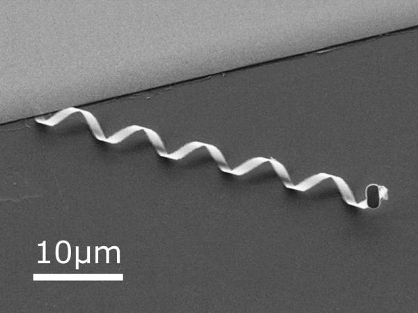
Made by depositing vaporized indium, gallium, arsenic and chromium onto a surface substrate a few atoms thick, the ABFs are patterned using lithography and etching. When thin sliced they naturally form the curled ribbon corkscrew you see in the picture above. The ABF head seen on the right contains a tri-layer film of chromium, nickel and gold. Nickel’s magnetic properties make it possible to use an external magnetic field to provide locomotion. The ABF uses helical propulsion (the same method of locomotion used by many bacterium) to swim through liquid at speeds of up to 20 nanometers at present with plans to increase it to 100 nanometers. Compare that to E. coli which swims at 30 nanometers per second.
To give ABFs autonomous power researchers are looking at thin-film rechargeable batteries. Currently these batteries can be manufactured and shaped with thicknesses of less than 50 nanometers. Chemical fuel sources may prove to be even more promising. An ABF running on energy it harvests from the blood would be capable of running indefinitely within a body harvesting glucose and oxygen to provide motive and computing power. Some researchers are looking at using bacteria as an ABF, modifying a living cell by attaching nanoparticles to it so that it can be mobilized for biomedical purposes.
What can ABFs do that current biomedical technology cannot?
Currently ABFs are confined to the research laboratory. But soon we will microscopically see these devices applied to all kinds of medical procedures reducing the need for surgical procedures, minimizing the side affects of cancer chemotherapy, and giving biomedical professionals a new set of tools for tackling conditions for which current medical practice has few solutions.
For those developing this technology it’s a question of tinkering with the power source to come up with the best way to deploy micro-robots.
]]>In unlocking the mysteries of the human genome we are learning to master the very essence of what makes us human, that orders and sequences our anatomy and operates our physiology, that explains why some of us are more susceptible to particular diseases than others, that tells us who is likely to get cancer, develop early onset Alzheimer’s Disease, or pass a congenital illness to our descendants.
DNA is the programming language of life. It has been in existence on Earth for almost 4 billion years. In the last decade of the 20th century and the first of the 21st we have mapped our genome in its entirety. We in effect are in a position to replicate the genome and even make improved copies of ourselves. The science we have practiced in agriculture we are starting to apply to ourselves.

Today genetic sequencing is the fastest growing biomedical industry on the planet. Biology has become biotechnology with ever more powerful computers central to us building new proteins, medicines, and biology. We are writing genetic code just as if it were a programming language. Instead of writing software we now are writing life. We have synthesized viruses and bacteria. When will we be capable of “creating” multicellular life? We are not far way. For those who profess religious faith they would say we are playing at being God.
The truth is we still do not know how life got started on Earth. So we don’t know how nature made us but we have discovered the tool kit nature uses. We have also looked into ourselves and made other discoveries. We are not a single entity. We are in fact a combination of 10 trillion cells and a host for 100 trillion bacteria and viruses that help us thrive. In knowing our genome we have the means to engineer the 10 trillion cells. But we can also alter ourselves bacterially to improve our health and prolong life.
In 1900 we were humanity 1.0. Understanding our biology remained in its infancy. Insulin for treating diabetes was only discovered in 1921. The first natural antibiotic, penicillin, was identified in 1928. These two medical discoveries can be linked to the development of humanity 1.1, a species interdependent with the medical support system that evolved in the 20th century.
In our mastery of the DNA toolkit what will humanity 2.0 be in 2050, and what will 3.0 be in 2100?

Here are some of the issues and technology trends that will drive biomedical innovation in the 21st century:
To learn more about each of these topics revisit this blog in coming weeks as we tackle each individually. As always, questions and comments are highly welcome.
]]>


