Before surgery, I had been dealing with osteoarthritis in both knees for almost two decades. My left knee was far worse than the right. I had been receiving cortisone injections regularly to keep me ambulatory. But in July 2020 I had a prosthetic joint implanted.
For years I had put off the surgery in the hope that a less invasive therapy using stem-cell-generated cartilage would become available. It is cartilage that provides cushioning to allow the knee joint to work without bone-on-bone contact. In osteoarthritis, the cartilage wears away until nothing is cushioning the immense stresses and loads that knees take on during daily activities.
As a surgeon explained to me, the reason stem-cell therapies to repair knees has yet to succeed isn’t that we cannot generate cartilage from stem cells, it is the collagen found in knee joints that cannot be restored or regrown. You may be familiar with collagen because of the many beauty supplements and treatments being sold on the market today. The collagen used for these isn’t the same as collagen found in knee joints. The complex collagen proteins in the knee are very different and play an important role in cartilage maintenance. Although we can generate chondrocytes (cartilage from stem cells), producing collagen suitable for knee therapies this way has yet to be done successfully.
So what research into non-surgical alternatives is being worked on today? An article entitled “Adhesive hydrogels in osteoarthritis: from design to application” appeared in the Springer Nature journal, Military Medical Research on January 30, 2023. It described research into developing hydrogels for osteoarthritic knee joint repair. Either natural or synthetic, the adhesive characteristics of hydrogels could be an answer.
As previously stated, hydrogels have great adhesive characteristics making them a good collagen substitute when attempting to regrow and restore cartilage in knee joints. The hydrogel material infiltrates the pores and irregularities found on tissue surfaces. This characteristic is called mechanical interlocking and it produces flat, slippery and firm surfaces. Injecting a layer of chondrocytes after hydrogels have bonded with the knee joint’s bone surfaces could become a potential non-surgical treatment. The problem, however, is that although the hydrogels bond to the bone, so far, getting the cartilage cells to do this has been another story.
A range of different adhesive hydrogels is being evaluated. The ideal is a hydrogel with double-sided adhesive characteristics to bond both bone and cartilage. Some are composed of contrasting structures. These are referred to as having double-network characteristics, elasticity on one side and stiffness on the other. Current research is looking at bacterially-engineered hydrogels coming from natural sources such as barnacles, mussels and sandcastle worms.
A material challenge for hydrogels will need to be tested when hardening. When used on knees they cannot be too brittle or too soft. The hydrogels have to stand up to the humid environment that characterizes our knees and bodies (we are mostly water). They have to respond to wear and tear caused by enormous stresses put on knee joints. And they have to be applied to the joint in the least invasive way possible. That’s a lot of issues still to be sorted.
The authors, in concluding their paper, summarize what future hydrogel-based treatments must provide:
When I started my search for an alternative treatment for my osteoarthritis, it was more than a decade ago as I read and learned about stem cells, chondrocytes and collagen. And it likely will be another decade before non-surgical knee repairs using the techniques and materials described above become mainstream. The solution needs a multidisciplinary approach involving chemistry, biology, pharmaceuticals and significant clinical trials.
]]>In the past transplanted stem cells have exhibited very low survival rates. But Dr. Karimi explains what’s novel about the work being done at the University.
“We have discovered that presence of a class of inhibitory molecules in the injured spinal cord potently limits success of stem cell transplantation. When we blocked these molecules therapeutically at the time of transplantation, the treatment led to an increase in stem cell survival and generated suitable neural cells in the injured spine…This strategy enabled rats to walk with more weight support and coordination.”
I have a friend who suffered a traumatic spinal cord injury (SCI) in his teenage years and has been confined to a wheelchair ever since. The health and work challenges he has faced would make the average person curl up in a ball and check out of life. But Dan has forged ahead and created a business to help others with disabilities. He has even coached wheelchair basketball teams in the past.
In Dr. Karimi’s laboratory, the work has been led by a doctoral student, Dr. Seyed Mojtaba Hosseini, who is the lead author of a paper describing the research and results, recently published in the Journal of Neuroscience.
The paper states that SCI “is a leading cause of permanent neurologic disabilities in young adults. Functional impairments after SCI are substantially attributed to progressive neurodegeneration.”
According to the World Health Organization (WHO), between 250,000 and a half million annually suffer SCIs. The majority are caused by traffic accidents, falls or violent confrontations. The WHO notes that those who suffer from SCIs “are two to five times more likely to die prematurely than people without a spinal cord injury.” In addition, SCI victims have “lower rates of school enrollment and economic participation,” and low employment and earnings while enduring significantly higher costs of living.
At the University of Manitoba the researchers identified two receptor proteins (molecules) that inhibited the regeneration of motor and spinal interneurons. By suppressing these proteins in the laboratory in female rats, transplanted neural precursor cells (NPCs) were able to thrive and replace damaged neurons. If these results can be repeated in human patients, the treatment strategy could prove promising for neuronal replacement in SCIs.
In Canada, in 2019, the annual cost was estimated to be $2.7 billion to cover medical management, hospitalization, and indirect costs. Long-term issues like low employment and subsidies were not factored in the data.
From U.S. data for the same year come the following numbers:
There is no doubt, that finding a therapy to get those with SCIs back on their feet literally would dramatically reduce lifetime costs for care, as well as give those with SCIs better prospects at long-term employment.
Dr. Karimi in the University of Manitoba press release talks about overcoming the consequences of SCIs. She states,
“Individuals with spinal cord injury live with the debilitating consequences of the disease for decades, and development of new regenerative medicine treatments is an unmet need to improve the quality of life for a large population of people.”
Further study of this approach to treatment is warranted and Dr. Karimi hopes to proceed to human clinical trials in the future.
]]>

Because stem cells are being heralded as the shape-shifters within our bodies, a universal panacea to cure almost everything that ails us. Biopharma companies recognize the money opportunity and are starting to bank them. Seen as the newest biopharmaceutical growth industry as opportunities to create new wonder drugs are narrowing, and as time runs out on exclusivity for so many of the drugs used globally, personal medical cures are all the rage. According to Market Reports World, analysts predict annual growth of stem cell banking at 12.76% from 2018 to 2022.
What are the market drivers?
Where are stem cells being used in medicine today? By no means is the following a complete list but it is a good starting point.
How legitimate are these stem cell therapies?
Some show great promise. Most recently, clinical trials using stem cells have created hope, not hype, for cures with patients suffering from childhood leukemia, sickle cell anemia, and dry macular degeneration. All of these stem cell treatments have come after more than a decade of animal studies followed by small-scale human clinical trials. In one example involving blindness from the death of rods and cones in the eye’s retina, transplanted stem cells repurposed themselves to replace the dying cells leading to the restimulation of optic nerve activity. These were stem cells harvested from the patient themselves and then cultured to increase their numbers before being injected into the retina.
Who are the players in the stem cell banking and treatment game, and where are they located? This list comes from Market Reports World with my added annotations:
It remains unclear just how effective stem cell therapies will ultimately be. But there is no doubt that the use of these undifferentiated shape-shifters is altering the way numerous diseases are being treated. I have copied a table that appeared in an article entitled Umbilical Cord Blood: Information for Childbirth Educators. It was written and posted to The Journal of Perinatal Education in its winter 2011 issue. The list appearing below represents what was seen then as the potential uses of stem cell therapies in tackling a range of diseases.

]]>
What are mesenchymal stem cells?
Known by the acronym MSC, these are cells found in our bodies. They are the undifferentiated source of the stromal cells in our bodies, the cells that form all of our connective tissue from organs to the lining of our gut. They can differentiate into bone cells called osteoblasts, cartilage cells called chondrocytes, muscle cells, called myocytes, and fat cells, adipocytes. They are referred to as multipotent because they can differentiate into all these different types of tissue. That’s what makes them so interesting to the field of study known as regenerative medicine.
Scientists have always looked at salamanders and lizards with wonder. How do these creatures drop a tail to a predator as a defense mechanism and then regrow it? Are stem cells involved?
In both cases, the answer is yes. Tail regeneration in both animal species happens when genes send instructions to the stem cells located in proximity to the severed appendage which signals differentiation to begin.
Differentiation is a fascinating aspect of living creatures. An apt analogy is to think of DNA as a movie script with the specific genetic information leading to differentiation, an individual actor’s part. When the actor reads the lines of the script within a scene, those words spoken are akin to the instructions delivered to proteins responsible for transcribing genetic messages to switch on cell machinery that makes an MSC begin to differentiate during cell division into new cell types.

Do humans share the same genes common to lizards and salamanders that can help regrow lost tissue?
Since we share so much of the same DNA, the answer is yes. But in our species, the switch to turn the genes on tends to be restricted or in the off position most of the time. So modern medicine is seeking ways to provide additional stimulus to kickstart regenerative processes. Harvesting our own MSCs appears to be one way to heal ourselves much the way salamanders and lizards do.
There are two ways to harvest our own MSCs. One is to take a small quantity of the cells and then culture them in a laboratory until you have a significant population. The second is to harvest the cells from our bodies where they can be found in large quantity so that they can be immediately be used in a medical procedure. The MSCs, in either case, are injected into an area of the body where there is damaged tissue and then stimulated to differentiate.
So where do you go in the body to find a significant quantity of MSCs?
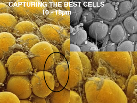
If you answered bone marrow, you are wrong. The answer is fat or adipose tissue which has 500 to 1,000 times the concentration of stem cells compared to bone marrow. Adipose-derived regenerative cells are today being used in clinical trials here in North America. Worldwide more than 10,000 patients have undergone regenerative medical treatment using their own MSCs. Here in Canada to date, there have been no clinical trials using MSCs. Canada’s only trials have used embryonic stem cells.
What is wrong with just doing trials with embryonic stem cells?
Embryonic stem cells, as promising as they have been, must be cultured in laboratories to grow a uniform population of sufficient volume for transplantation. What guarantees their genetic stability? The laboratory protocols covering generation and maintenance of cell populations is critical to ensuring that all embryonic stem cells function normally after transplantation without the potential for a mutation occurring in one or more of the cells. A mutation could lead to unintended consequences such as tumors with some potentially malignant.
Embryonic stem cells, when used in treatment, can be derived from the patient but the process to create them involves extracting pluripotent adult stem cells and reverting them to their embryonic state before culturing them to grow a population. Embryonic stem cells can also originate from a third party’s adult stem cells which through reversion can then become useful for regenerative therapies.
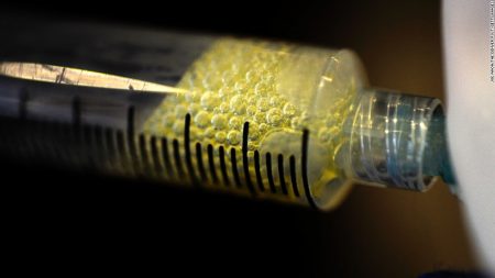
MSCs used in regenerative medicine procedures don’t require the complexity that is characteristic in using embryonic stem cells for therapy. They can be harvested from adipose tissue, our body fat, in large volumes, sufficient for therapeutic purposes and can be done with one particular technology (you’ll have to read Part 4 to find out which) in a single minimally invasive treatment process, done under local anesthetic, that involves three steps.
So what makes the transplanted MSCs differentiate?
With all cells sharing the same DNA it seems locality determines the type of differentiation that follows. So if you inject MSCs into a knee joint the MSCs will differentiate into chondrocytes. Put them into a liver and they will differentiate into the tissues found in that organ. Put them into a heart, or muscle and voila you will see differentiation produce the desired tissue.
How long does it take before results occur?
Differentiation begins almost immediately as cells replicate. But how does that translate in terms of time before the patient sees improvements? Depending on the medical issue a patient may begin to see improvements within a few days although with some conditions it may take up to six months for the regenerative treatment to work largely dependent on tissue type and normal replication rates through cell division.
In Part 4 of this series on regenerative medicine, I will introduce a technology that may prove to be a game-changer for regenerative medicine. Today, it is being used in a series of clinical trials around the world and its inventors are based in Richmond, British Columbia.
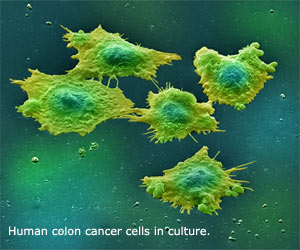
The research at Princess Margaret has specifically focused on colon cancer and the role BMI-1 plays. What they managed to do is disarm the BMI-1 gene to stop colon cancer stem cells (see image below) from creating a new tumor. They did this by removing colon cancer cells including cancerous stem cells from patients with the disease and exposing the cultures to a new drug BMI-1 gene inhibiting drug that is still being developed and tested. The results – the cancer stem cells were disabled.
Cells from the same cancerous cell culture were also injected into mice specifically bred to tolerate human tissue transplants. The mice grew tumors within a few days. The drug treated cells were then injected into the mice, and then remnants of tumors in these mice were extracted and transplanted into a second group of mice. The results again showed a dramatic drop in cancer stem cells. The results of the study were published on the weekend in Nature Medicine.
For those in the medical field fighting cancer these findings provide a therapy that may permanently end a malignancy eliminating recurrence. The BMI-1 biomarker appears in 65% of patients with colon cancer. By inhibiting self-renewal of cancer cells in patients with the biomarker the recurrence of the disease which happens in about 50% of patients can be eliminated.
It is still too early for clinical trials but BMI-1 cancer treating drugs should be on the market within the next ten years and this research is the first step in getting us there.