| July 7, 2020 – In the latest missive from Peter Diamandis, he focuses on advances in our medical and scientific knowledge around stem cells, organ generation, bioprinting, and extending human lifespan. Peter has picked an interesting time to be full of optimism about the future of our species as we face a current pandemic and the mounting threat of anthropogenic atmospheric and ocean warming. He justifies this optimism because he sees disruptive forces of innovation breaking the back of our current problems, and is looking to a far brighter future for the planet and us. Here are mostly his words and ideas which I have edited where warranted to share with my readers. I hope you find the subject intriguing and maybe even a bit disturbing. As always your comments are appreciated.
Lizards can regrow entire limbs. Flatworms, starfish, and sea cucumbers can regrow entire bodies. Sharks replace lost teeth, as many as 20,000 throughout a lifespan. So why can’t we do the same? The answer: Through cutting-edge innovations in regenerative medicine, research is underway to do just that. In 2019, investors pumped $10 billion into this field, and currently, there are over 1,000 clinical trials underway trying regenerative treatments on 60,000 patients. As Big Data and Artificial Intelligence (AI) transform medicine, regenerative medicine will take us to these three Rs:
1. ReplenishStem cells are undifferentiated cells that transform into specialized cells from embryos to when we are born and beyond. They can be found in many areas of the body. Stem cells differentiate to become heart, neurons, liver, lung, skin and other specialized tissues. They also can divide and produce more stem cells. As a child or young adult, we have stem cells in large supply. They trigger when the body needs to repair itself. They are summoned to sites where there is damage or inflammation. As we age, the supply of stem cells diminishes by as much as 100- to 10,000-fold throughout different tissues and organs. Stem cells also undergo genetic mutations over a lifespan which reduces their quality and effectiveness at repairing damage. If you could restore and rejuvenate a body’s stem cell population we can extend our healthy years dramatically. One way to do this would be to extract and concentrate our autologous adult stem cells harvested from adipose (fat) tissue or bone marrow. Another source comes from umbilical cords and placentas kept after birth. The advantage of the latter over the former is these stem cells have not been subject to aging like those found in adult adipose tissue or bone marrow. Umbilical and placenta stem cells provide the undamaged software of a newborn and when cultured to multiply, can be injected into joints or administered intravenously as a rejuvenation therapy. Using these stem cells can help to fight inflammation, autoimmune disease, increase muscle mass, repair joints, and even revitalize skin and grow hair. A burgeoning field, stem cell research has seen a 40-fold increase in scientific publications over the last few years. The market globally is estimated to grow to $14.8 billion USD by 2022, growing at an annual growth rate of 14.7%. Examples of regenerative treatments include:
2. ReplaceEvery 10 minutes an American is added to an organ transplant waiting list. As of March 2020, 112,000 people were waiting for replacement organs. Countless more people never make it to the waitlist with an average of 20 dying each day. The result, 35% or approximately 900,000 people can be prevented from dying with better and more timely access to replacement organs. Promising a remedy to organ shortages, replacement and regenerative medicine represents a massive opportunity. The opportunity has led to emerging organ entrepreneurs. One of these is United Therapeutics, started by Dr. Martine Rothblatt, a one-time aerospace entrepreneur and founder of Sirius Satellite Radio. Personal circumstances led to a change of careers when Rothblatt’s daughter developed a rare lung disease. Her “Moonshot” project to replace her daughter’s unhealthy lungs, and others with similar affliction, has become a major business opportunity pursuing three disruptive strategies that could change medical transplants forever. Rothblatt’s company is United Therapeutics where she is CEO. With an initial focus on diseases of the lung, she has set out to create replacement lungs. To accomplish this goal, she has pursued a number of technologies in parallel including. A. 3D Printed OrgansIn 2017 she teamed up with one of the world’s largest 3D printing companies, 3D Systems, to build a collagen bioprinter. She teamed up with another company, 3Scan, to create maps of the lungs structures and interior using detailed micro slices programmed into a bioprinter. An ultraviolet laser passing light through a shallow pool of collagen doped with photosensitive molecules served as the medium for the build. The laser targeted the areas identified by the mapping causing collagen to cure and form a scaffold to which human cells attach. So far the technology has been able to produce lung structures with resolutions of 20 micrometers. The end goal is to get to one micrometer to achieve a functioning replacement for a human lung. Once a scaffold capable of these dimensions can be built it will be infused with living stem cells using recellularization. Stem cells will grow and differentiate, populating the scaffolding, and producing a functional lung. Can it be done? In 2018, Harald Ott, an experimental surgeon at Harvard University reported that he had pumped billions of human cells (from umbilical cords and diced lungs) into a pig lung stripped of its own cells. When Ott’s team reconnected it to a pig’s circulation, the resulting organ displayed rudimentary lung function. B. XenotransplantationRothblatt is pursuing the transplanting of animal organs into humans which is known as xenotransplantation. Because an adult pig has organs similar in size and shape to those of humans, her company is focusing on genetically engineering pigs to harvest their organs for human transplants. “It’s actually not rocket science,” said Rothblatt in a 2015 TED talk. “It’s editing one gene after another.” To accomplish this goal, her company has made investments in Revivicor Inc. and Synthetic Genomics Inc. signing funding agreements with the Universities of Maryland, and Alabama, and New York-Presbyterian/Columbia University Medical Center to start a xenotransplantation program for hearts, kidneys and lungs. Rothblatt hopes to see the final results in three to four years. The end goal for the company will be to produce up to 1,000 sets of healthy pig lungs, called xenolungs, annually from genetically engineered pigs. C. Ex Vivo PerfusionToday only 30% of human-donated lungs meet transplant criteria. Of these, 85% remain usable upon arrival at a surgical site for implantation. The end result, nearly 75% of donated lung tissues never get used by recipients in need of a transplant. But what if the donated lung tissue could be rejuvenated? In 2016, Rothblatt invested $41.8 million in TransMedics Inc., an Andover, Massachusetts company that has developed the XVIVO Perfusion System which takes marginal-quality lungs initially fail to meet transplantation standard-of-care criteria and perfuses and ventilates them in normothermic conditions, to provide an opportunity for surgeons to reassess their transplant suitability. The perfusion system can also be used with heart, kidney, and liver tissue for transplantation. RejuvenateResearchers at Stanford and Harvard University have demonstrated that older animals when transfused with blood from young ones experience regeneration in many of their tissues and organs. The opposite is true as well that when young animals are transfused with blood from older donors, they experience accelerated aging. Elevian (a Harvard University spinoff) is using this “young blood” effect in its approach to studying longevity. CEO Mark Allen, MD, is among the founders along with a dozen MDs and PhDs from the university. They have identified specific factors responsible for the effect described in the previous paragraph. A naturally occurring molecule known as “growth differentiation factor 11,” or GDF11, which, when injected into older mice, reproduces regenerative effects in the heart, brain, muscles, lungs, and kidneys. Further, GDF11 reduces age-related cardiac hypertrophy, accelerates skeletal muscle repair, boosts exercise capacity, increases brain function and cerebral blood flow, and even improves metabolism. From this Elevian is developing a number of therapeutics to regulate GDF11 and other circulating factors with a goal to restore the body’s natural regenerative capacity. The company believes with its current research it can counter the root causes of age-associated disease promising to reverse or prevent many of them and to extend a healthy lifespan. Final ThoughtsIn 1992, the futurist Leland Kaiser created the term regenerative medicine. He called it “A new branch of medicine…that attempts to change the course of chronic disease and in many instances will regenerate tired and failing organ systems.” From these initial roots has grown an industry that in 2018 generated $23.8 billion USD and is projected to exceed $151 billion by 2026. If the work described here pans out, the road ahead for improvements to human health will be enormous. Imagine having the ability to regenerate, replenish, and replace entire organs and metabolic systems on command. Are there unintended consequences to Peter’s hopeful description of the biomedical breakthroughs that lie ahead? Extending the natural lifespan and making for a longer and healthier lives in a person’s 70s, 80s, 90s, and even 100s and beyond comes with an added burden to the planet which already has 7.8 billion people on it. What are the implications for work and retirement? What are the implications for subsequent generations seeking the jobs of those who are older and no longer prepared to retire because of aging and disease? At the beginning of the 20th century, the average human lifespan was 40. By the beginning of the 21st century, it had almost doubled. By 2100, will it double again? Peter Diamandis is convinced it will and we are seeing in this article the beginnings of the technologies that will make that a reality. |
The concept of rejuvenating our bodies through the use of stem cells is no longer a foreign idea. It comes with a degree of controversy as clinics offering stem cell treatment are popping up across the world making promises to cure diseases, restore joints, ease arthritis, and cure cancer. That promise remains tantalizing because the idea to be able to regenerate any damaged tissue in you using cells from you seems fantastic.
Why is the promise of stem cells possible?
The human body is unbelievably smart. It can recognize foreign cells and organ rejection can occur even in cases when a donor is considered to be a near-perfect match to the patient. But grow an identical heart derived from the patient’s own cells, and the results may be different. So in a world where technology is evolving to accommodate anything’s possible, why not tissue regeneration.
When did we take the first regenerative medicine steps?
It first started in 1954 with the first kidney transplant. And today, according to the Alliance for Regenerative Medicine, there are 1,060 clinical trials and 900 companies globally focused on this field. Here’s an idea of what financing for the industry currently looks like:

Not quite. Regenerative is a subset of personalized medicine, an approach that attempts to tailor-fit treatment to an individual. It is a departure from the current practice by physicians and biomedical companies of “one-size-fits-all” medicine. It could mean tailoring or developing a unique drug treatment to match a person’s DNA. With regenerative medicine the treatment is designed to allow the body to repair itself through cell, tissue, and organ replacement. The treatments are hyper-customized using a patient’s own stem cells. As you can imagine, getting this type of treatment to scale is proving to be pretty expensive. But its promise has lured close to one thousand companies to work in the field as can be seen in the graphic displayed below.

The Alliance for Regenerative Medicine has compiled the clinical trials by medical category. The largest number are devoted to oncology followed by heart and circulatory diseases, diseases of the central nervous system, and musculoskeletal diseases. A visual breakdown of trials can be seen in the image below:

Treatments currently on the leading edge include:
As exciting as this burgeoning field is, the high cost involved and the cutting-edge nature of the technology is holding the field back. But the data shows that things are looking up. Research and Markets predicts a staggering 23.35% compound annual growth by 2022 compared to 2016 numbers. And experts forecast growth to $66 billion annually.
Why not? Lowered risks, minimally invasive procedures, and more efficient disease treatment and prevention should eventually guarantee lower costs and higher recovery and cure rates. Just what insurers, hospitals, and doctors want.
]]>I have a personal interest in MSC for use in treating osteoarthritis, something I have suffered from for more than 45 years. I have always been concerned about the viability of stem cell injections particularly in terms of rates of survival once transplanted into an injury site. One reason for concern is how the cells will defend themselves from the body’s natural immune system. MSC implants could be subject to attack from T-cells and other white cells that would treat their arrival as a foreign substance.
But what Ghosh and her colleagues have created is a protective transporting hydrogel shell that contains the MSC supporting the viability of the cells for as much as a month after injection. The hydrogel is a concoction of cellulose-like material and chitosan, a derivative of chitin which forms the exoskeleton of crustaceans like shrimp.
In an article appearing in yesterday’s The Hindu Business Line, Ghosh is quoted stating, “the hydrogel addresses the issue of the long- term survival of adult stem cells in simulated cultures that mimic the actual body tissue. We observed that cells survive and multiply for a period of one month, which is sufficient time for tissue regrowth.” In other words, the hydrogel doesn’t interfere with the capability of the MSC to interact with neighbouring cells and stimulate division.
Ghosh’s hydrogel, following implantation, supports normal functioning of the MSC contained within it. Subsequently the MSC secrete a growth factor called paracrine which signals nearby non-stem cells to alter behaviours. MSC injected into a knee joint protected by the Ghosh hydrogel would, as a result, in no way inhibit signaling to chondrocytes, cartilage cells, or osteocytes, bone cells, to begin to multiply. It is interesting that the two cells tested by Ghosh and her team were chondrocytes, and fibroblasts, the latter skin cells, and showed no inhibiting factors. The next step will be to apply what they have learned in the laboratory to animal studies. And if those are successful we should see human clinical trials in the near future.
The potential of the hydrogel-MSC composite injection serving as scaffolding for reconstructing knee cartilage or repairing wounds and damaged organs cannot be underestimated. The injectable’s ability to take the required shape within an injection site and provide adhesion to the surrounding tissue is a significant breakthrough that if replicable in animal and subsequent human patients, could change the way osteoarthritic joints and other injuries get treated in the near future.

I have been looking at knee replacement surgery for the last ten years and resisting the idea of going that route in the hope that stem cell therapies and regenerative technologies advance to the point where they represent a viable option. If I could regrow the cartilage in the knee joint to the point where it gives me pain relief it would seem to be the best of all solutions.
There are clinics operating in Toronto today that offer knee injections using harvested mesenchymal stem cells taken from my own body fat. The harvested cells, being my own, would not be rejected. And in proximity to the joint might start growing chondrocytes – cartilage cells. The question I have always posed to anyone offering this therapy is “how do the cells know where to grow and when to stop populating the joint?” To date, none of the clinics have given me a satisfactory answer.
In my research, I have noted that some universities are experimenting with gel-based scaffoldings which can be implanted in a joint and become a seedbed for regrowing chondrocytes. The scaffoldings over time would dissolve leaving the repopulated cartilage in place. That’s why when I saw the announcement today for a call for papers by Drs. Francesca Diomede and Jacopo Pizzicannella, of the University “G. d’Annunzio” Chieti and Pescara, in Italy, I was elated. The call for papers includes a covering note which states, “The use of a biofunctionalized scaffold with cells and/or soluble factors has emerged as a promising approach in the field of regenerative medicine.” It goes on to describe what is meant by a biomaterial, an environment that supports the growth and development of cells allowing them to adhere and proliferate. Although the letter specifically addresses bone regeneration, the goal of being able to create a therapeutic alternative to traditional surgical intervention should equally apply to the regrowing of cartilage in a biofunctionalized scaffold designed to dissolve over time.
The deadline for manuscript submissions is December 31, 2019. I’m happy to hang on for another year and see where regenerative medicine is going to take us. The last thing I want to consider is an artificial knee joint.

]]>
ESA has been working on health and survivability issues for astronauts going on Deep Space missions. Whereas an astronaut or cosmonaut on the International Space Station can return to Earth in the event of an onboard medical emergency, those undergoing long-duration missions to the Moon, Mars, or a future asteroid visit will be too far away for medical intervention in the face of a serious injury.
ESA is also looking at bioprinting and regenerative medicine as a means to counter the effects of prolonged exposure to cosmic radiation, and to the physiological impacts to the body in low-gravity environments such as Mars and the Moon. The idea is to be able to print skin, bones, and organs.
The missions ESA is evaluating for the use of bioprinting and regenerative technologies are long-duration including journeys to the Moon with stays lasting weeks or months, or an exploratory mission to Mars with human crews in interplanetary space for six months or more en route and on the surface of the planet for a month or more.
Among the materials that the ESA researchers are studying are hydrogels which are thermoplastic polyurethanes that can create scaffolding to repair and regenerate load-bearing soft tissues like cartilage, tendons, and blood vessels. Hydrogels are great for regenerative medicine because they can be easily extruded through a 3D printer, and are water friendly which makes them a perfect medium for restoring and regenerating soft tissues.
The use of 3D bioprinting today is still at the early stages of development. Today the best that 3D printers can do is manufacture prosthetics for legs and arms, create laboratory-generated tissues that have the characteristics of human organs on a minuscule scale, and build accurate models of bone and tissue that surgeons can study prior to doing complex procedures. These 3D printers get their data from diagnostic imaging technologies which render digital images and data that can be interpreted. But it takes considerable time to render the digital images into detailed printing instructions, sometimes days. So the next advancement to get to where ESA wants to go is to take computer-generated radiology images of bones, blood vessels, and organs and using software generate precise printing instructions in a matter of minutes once an injury has been assessed.
Of course, what ESA wants to deliver in Deep Space will have profound and meaningful use here on Earth. The field of replacement therapy and regenerative medicine using 3D bioprinting will alter how medicine is practiced. Imagine replacing a defective kidney with a healthy 3D printed one ending the need for dialysis. Or replacing defective hearts, lungs, livers, bladders, ovaries, uteruses, pancreases and other body parts eliminating much of what medicine has to treat today.
When will 3D bioprinting of replacement body tissue become a reality? Sometime within the next decade believe the experts in this very new technological field.

]]>
Recently the FDA approved a number of gene therapy products to treat childhood and young adult leukemia, non-Hodgkin lymphoma, and forms of congenital blindness. The guidelines for giving the green light to Novartis, Kite Pharma and Spark Therapeutics, the companies responsible for producing these new genetic treatment protocols are covered in a new document entitled, “Chemistry, Manufacturing, and Control Information for Human Gene Therapy Investigational New Drug Applications.”
Gene therapies involve the transfer of healthy genetic material into gene sequences to replace bad genes responsible for a particular medical condition. Regenerative medicine involves the therapeutic re-engineering and regrowth of cellular tissue of all types from organs, to bones, to nerves, blood vessels, and the like.
In announcing the new guidelines an FDA spokesperson and health policy expert described how different these new therapies are from traditional drug development and treatment protocols. Whereas the focus in the latter is 80% on clinical issues, when it comes to gene therapies and regenerative medicine the 80% is on manufacturing looking at:
The FDA also has included long-term follow-up guidance for gene therapy and regenerative medicine products. The Agency admits it is still on a learning curve to understand how these new therapies work and what is considered safe in the long term. But in the interest of initial results from laboratory and clinical trials, the goal is to approach these forms of medical treatment with an evolving strategy based on the rapid evolution of the science. So expect to see frequent revisions to FDA treatment protocols as new genetic and regenerative medical therapies are announced by biopharmaceutical companies and academic researchers.
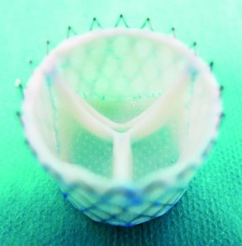
The effort is part of the European Union-funded LifeValve project, aimed at replacing mechanical and preserved animal tissue valves which are the current standard. The medical team cultures the heart valves from human cells. Design of the valves is done using computer simulations based on detailed imaging of a patient’s heart and outflow tracts to ensure that what is regenerated can be implanted without fitting problems.
Initially, the team used computer simulations to help understand how to culture human cells and grow them into a heart valve. They chose sheep for their animal models and were able over time to come up with accurate bio-engineered valves that could be implanted successfully. And now they have done it with the first successful customized implants in living sheep.
Why is this development so significant?
On a personal note, my daughter was born with a complex congenital heart defect called Tetralogy of Fallot with Absent Pulmonary Valve. She has had two open-heart operations and a third procedure where a valve was inserted into her pulmonary artery via a catheter. All three valves were cryogenically preserved dead animal valve tissue. My daughter faces a lifetime of these periodic valve replacements because dead tissue cannot regenerate itself.
Worldwide the incidence of congenital (meaning born with) heart disease (CHD) is 8 to 12 for every one thousand live births, amounting to over 1.3 million globally each year. This worldwide phenomenon knows no boundaries occurring in populations everywhere with only minor variation by country in CHD type.
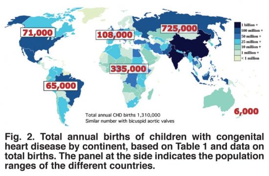
Structural repairs for most CHDs involves patching holes in the heart, altering arterial structures to improve blood flow either through the lungs or to the bodies extremities, and replacing missing or damaged valves. The human heart has four and depending on the type of CHD, any one or more of these valves could require a replacement implant.
With current practice using dead animal tissue valves, CHD patients undergo repeated surgical interventions throughout their lives. That’s because the valve flaps wear out, or calcify to become less flexible and restrict arterial blood flow. The alternative surgical prosthetic, mechanical valve implants, may last a lifetime, but require CHD patients to stay continuously on anticoagulants to prevent blood clots. But growing a heart valve with the CHD patient’s own human cells would eliminate the problems attendant to both current practices.
The biggest challenge is no longer in the growing but in the custom fitting of what is grown. There is no one size fits all solution. But it appears that the work being done in Zurich may soon be reflected in the first human clinical trials giving children born with congenital heart disease a much better life requiring fewer surgical interventions. Eventually, the bio-engineering of heart tissue using a patient’s own cells could heal the hearts of all children with CHD.
The results from Zurich have been published in the journal, Science Translational Medicine, May 9, 2018 issue, in an article entitled, “Computational modeling guides tissue-engineered heart valve design for long-term in vivo performance in a translational sheep model.”

June 3, 2018 – I don’t know about you but going for my semi-annual dental checkup is not one of my favourite pastimes. When I give someone rights of access to my mouth I can’t help but remember the movie “Marathon Man” and the nightmares it precipitated. Fortunately, I trust my dentist and am glad I chose her. But what will I tell her when I see her this month after reading about the latest new research from Queen Mary University of London, where scientists and engineers may have discovered a way to grow mineralized materials including tooth enamel and bone.
Tooth enamel is considered the hardest tissue in our bodies. And although bone is hard it can do one thing enamel can’t. Break a bone and the body can regrow. Break a tooth and you are out of luck. Once tooth enamel goes we have to have dentists deal with the consequences: cavities, pain, damage to the root, and eventually root canal and a crown. These are problems that more than half of the world’s population deal with in a lifetime, and the reason why dentists exist.
Sherif Elsharkaway, a dentist, and an author of the paper appearing in Nature Communications entitled, “Protein disorder-order interplay to guide the growth of hierarchical mineralized structures,” describes the work as “exciting because the simplicity and versatility of the mineralization platform opens up opportunities to treat and regenerate dental tissues.” The mechanism behind regrowth is creating hierarchically organized architecture from apatite nanoparticles that mimic the characteristics of natural human enamel. Controlling the mineralization opens up opportunities for regenerative medicine including regrowing tooth enamel.
The lead author, Professor Alvaro Mata, Queen Mary’s School of Engineering and Materials Science, describes the utility of the discovery in terms of our ability to learn from nature. He notes that being able to have precise control at the molecular level makes it possible “to exploit disordered proteins to control and guide the process of mineralization at multiple scales…..a technique to easily grow synthetic materials that emulate such hierarchically organized architecture over large areas and with the capacity to tune their properties.”
With my upcoming visit to the dentist in another two weeks, I can hardly wait to tell her that she may soon be practicing a very different type of dentistry.

In Part 1 of this series on searching for a better knee, I talked about my recent visit to a Toronto clinic where I was assessed and provided with exercises and a plan to improve my mobility related to my osteoarthritic knee. It was reassuring not to be talking about scheduling knee replacement surgery, a procedure which I have been avoiding for many years.
In talking to the medical team at the clinic I mentioned regenerative medicine and the use of stem cells as an alternative to an artificial joint. It appeared that I knew more about this subject than the people advising me. In some respect, this seemed surprising to me. But then I realized, if the focus of this clinic’s efforts was on rehabilitation, why would it know about regenerative medicine related to knee joints. After all, the clinic was overseen by a team of orthopaedic surgeons. But not all orthopaedic surgeons are just thinking cut and replace these days when it comes to osteoarthritic knees.
On Friday I was sent a press release from PR Newswire featuring Kevin Plancher, an orthopaedic surgeon who in his practice is looking at both surgical and nonsurgical treatment of hip and knee joints. In the release, it describes the 50-year history of surgical joint replacement in the United States and notes that according to the Mayo Clinic, there are 1.5 times more Americans living with hip and knee replacement than there are living with heart failure. It went on to state that the outcomes for the vast majority have been good. But it also noted that some patients end up with multiple replacements or continue to suffer pain and limited mobility even after replacement.
Also in the release, Dr. Plancher describes the progress being made using growth factors and stem cell therapy. This technology is minimally invasive involving injections into an affected joint. The release talks about animal studies that have been showing promise by reversing the breakdown of cartilage and accelerating healing.
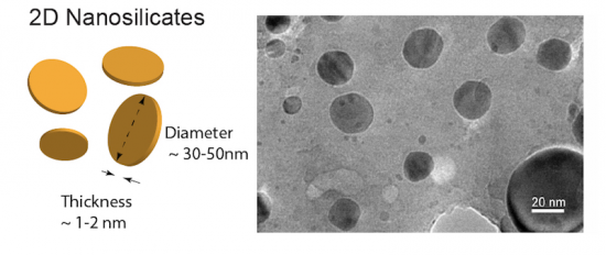
My own research into regenerative technologies indicates that there remain barriers to the use of stem cell therapy and it becoming an everyday procedure for restoring hips and knee joints. In the latest paper, I just read, however, researchers are introducing something new to regenerative therapy that could make the therapy viable. It is nanosilicates. What are nanosilicates? – a biodegradable synthetic material containing the mineral silica. When added to a bone grafting procedure, nanosilicate becomes a suitable scaffolding (like that seen in the picture below) that can be implanted into a joint to form the medium for regenerating bone or cartilage. Because nanosilicates are tiny they can put into a bio-ink containing a mixture of nanosilicate and stem cells that if injected into a damaged joint can begin the regenerative process.
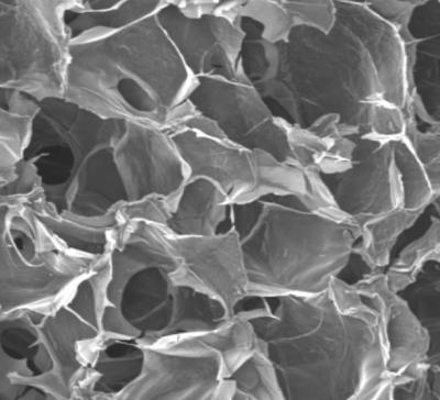
In the latest paper on the subject of regenerative medicine that appears in the U.S. journal, Proceedings of the National Academy of Sciences (PNAS), researchers at Texas A&M University, Sloan Kettering Cancer Center, and Weill Cornell, have described applying nanoparticles to grow bone and cartilage from mesenchymal stem cells from a human donor.
The team, led by Akhilesh Gaharwar, describe the use of two-dimensional nanosilicates, 30 to 50 nanometers in diameter and 1 to 2 nanometers thick, to provide a scaffolding for bone and cartilage regeneration therapy. The work being done by this team is part of a new field called mineralomics and could prove to be the breakthrough advancement needed to make stem cell therapies viable as a future treatment for knee and hip osteoarthritis.
States Gaharwar, “Osteoarthritis progressively degrades cartilage and often specifically attacks the spaces between joints. Because these intra-articular spaces lack blood vessels, systemic delivery of morphogens and growth factors supporting cartilage matrix synthesis usually results in limited improvement. These nanoparticles could be delivered to this space to stimulate chondrocytes to synthesize new cartilage matrix.” Although the current study described in the PNAS-published paper involved human cells in vitro, Gaharwar believes that the technology could conceivably be used “to develop implantable scaffolds containing these nanomaterials to improve local tissue growth.”
So it appears that computational biology, genomics, and next-generation bioactive materials like nanosilicates, combined with human stem cells may prove to be the breakthrough that turns regenerative knee and hip joints into an alternative to surgically implanted joints. And because the stem cells will be harvested from the patients themselves, the body’s autoimmune systems will accept the material without a negative reaction.
It got me thinking about some of the latest journal articles I’ve been reading on studies describing: how gene-edited pigs may soon become a primary source of organs for human transplants, about the recent reported genetic manipulation of an embryo’s DNA to correct a congenital heart defect, and about new stem cell research capable of regenerating organs, bone and even neurons.
Gene-Edited Pigs
The genetic tool CRISPR/Cas9 seems to be driving a biomedical revolution. In the case of genetically modified pigs becoming a source for harvesting organs for transplant, a new biotechnology company, eGenesis, is using the tool to modify the DNA of pigs to humanize their organs and eliminate rejection.
Animal organ transplants are known in the biomedical world as xenografts. In the past only inert, that is dead animal tissue, could be put into a human body. For example, my daughter’s first two implants were porcine xenografts, pig valves. The third was bovine, a cow valve. In all three the tissue was dead. The result, my daughter’s immune system did not react. If the valves had been alive then her immune system would have mounted an assault each time leading to rejection episodes.
Beyond the issue of foreign tissue rejection, there is an additional danger lying within xenografts. Within pig tissue lies viruses that are of little consequence to their porcine hosts. But if introduced into a human through a xenograft, can become a serious source of health problems. This issue plus rejection have been evident since the first efforts to use animal tissue in transplant procedures. So even if we were to manipulate a pig’s DNA to humanize it, the latent threat of endemic viruses might persist.
eGenesis is working on two remedies. The first involves manipulating pig cell DNA to humanize it so that when implanted in a human recipient it doesn’t set off the immune system. The second is working on pig cells DNA to eliminate native hidden viruses. The end result will be a merger of the two into a single cell and then use it to clone a humanized pig.
Will humanized pig organs be available for transplant soon? We are probably a decade away from seeing this become a reality. But it does make for great headlines.
The latest research is published in the journal Science in an article entitled, “Inactivation of porcine endogenous retrovirus in pigs using CRISPR-Cas9.”

Replacing Defective Genes
The latest headline on this subject appeared just a few weeks ago and I described it in an August 7th, 2017 posting on this site related to the repair of a faulty gene responsible for the congenital condition known as hypertrophic cardiomyopathy.
CRISPR/Cas9 appears to be the current tool of choice for altering DNA. It has been used for a number of years now to reverse disease symptoms in laboratory animals. It had not, prior to two recent experiments, been used with humans. But that is coming to an end particularly in helping prevent diseases caused by genetic disorders.
Genetic diseases are classified as monogenic, multifactorial or chromosomic.
In the case of the first, the genetic defect is confined to one gene. Examples of monogenetic disorders include sickle cell anemia, cystic fibrosis, Tay-Sachs, and polycystic kidney disease.
In the case of the second, it involves mutations in many genes. Heart disease, diabetes, and most cancers are multifactorial.
In the case of the third, it is associated with imperfections in the copying of chromosomes leading to abnormalities. The most well known of these is Down syndrome where an extra copy of the 21st chromosome occurs even though no genes within the extra copy are abnormal.
Microsurgery at the molecular level using a tool like CRISPR/Cas9, may prove to be the most significant advance in biomedicine in the 21st century. It could alter the aging process extending human life indefinitely. It could end cancer and many other diseases that today appear intractable. And on its dark side, it could lead to designer babies and super humans. How the consequences of this disruptive technology unfold remains unknown.

Stem Cells and Organ Regeneration
I have referred to stem cells, and particularly mesenchymal stem cells, as the “god cell” in previous postings on this site. That’s because mesenchymal stem cells (MSM) are undifferentiated cells hiding in plain sight within our body tissues. With the right stimuli, they can become bone, cartilage, muscle, and fat. It is this multipotential that makes these cells the key to the new field of study known as regenerative medicine.
Imagine regrowing a missing leg or arm. For a salamander that is no big deal. And for a male moose, elk or deer, regenerating antlers is an annual right of passage. But for humans regenerating body parts has been confined to regrowing muscle and bone after traumatic injury. In no case, however, has a human lost a leg and then regrown it, bone, muscle, cartilage, tendons, ligaments, toes and nails.
Manipulating stem cells to give humans the same abilities as a starfish or salamander is the dream of a number of scientists. Here are just some of their dreams being turned into realities:
And my favourite, at Duke University, a combination of 3D printing, hydrogels, and stem cells have been used to partially restore knee joints. And Duke is not alone in pursuing knee repair using a combination of stem cells and additive manufacturing. University of Gothenburg‘s Sahlgrenska Academy has been printing stem cells using a 3D printer to create cartilage which along with a cellulose matrix can restore an osteoarthritic knee, one just like mine.
Do we care which of these three biomedical breakthroughs wins out in the end? No. We need them all.


