Before I underwent knee replacement surgery three years ago last July, I was hellbent on waiting for a stem cell therapy alternative to regrow my arthritic joint. Having endured more than a decade of discomfort and a course of cortisone injections to keep the surgeons at bay, the advances in stem cell research had not yet reached a point where it could be considered a non-surgical alternative therapy.
There were several clinical trials. The closest still accepting volunteers was in Chicago. The only one in Toronto had reached its complement of candidates. Eventually, I was referred to an orthopaedic surgeon who explained to me why knee replacement using stem cells to regrow cartilage was still seeking answers for its most challenging limitation.
Mesenchymal stem cells harvested from a patient could be turned into chondrocytes, the name given to cartilage cells. Cartilage cells, however, need a matrix upon which to grow. That natural matrix is made of collagen, a structural protein our bodies produce that forms connective tissue. At the time of my referral, no stem cell treatment had yet to come up with a way to produce collagen and put it where it was needed to support growing cartilage.
The treatment of Liesel, the elderly gorilla, used mesenchymal stem cells harvested from a younger donor female gorilla who had undergone minor surgery in 2022. StemCellX, a company formed by leading stem cell scientists, worked with researchers from the University of Sheffield in the United Kingdom on Liesel’s treatment.
StemCellX is focused on cutting-edge research and stem cell treatments for veterinary medicine. It provided the harvested mesenchymal stem cells that were kept frozen until the procedure. There was no mention of concern about Liesel rejecting the stem cell infusion. That’s because mesenchymal cell transplants exhibit low rejection rates and tend to be well tolerated in transplantation. There was high confidence that Liesel would benefit since StemCellX has successfully treated dogs and horses with moderate to advanced osteoarthritis.
Several clinical trials using mesenchymal stem cell therapies for treating osteoarthritis have recently reported success. One from Brazil documents the use of bioscaffolds made up of collagen or hyaluronic acid applied to knee joints in place of natural collagen with intra-articular injections of mesenchymal stem cells to induce cartilage regeneration in osteoarthritic knee joints. The following illustration appears in the journal article that describes this clinical trial.

In another two-year case study published in the journal, Regenerative Medicine, 329 patients with knee osteoarthritis received mesenchymal stem cell therapy with significant improvements to the knee joint. In this clinical trial, the goal was to reduce or delay the need for total joint replacement.

In a University of California, Irvine (UC Irvine) clinical trial, laboratory-made neurons grown from embryonic stem cells have been injected into the brains of 12 patients with Parkinson’s. The clinical trial is ongoing but initial results are encouraging showing that the added neurons have formed synapses to link to the patients’ brain cells and produce dopamine.
Dopamine is a neurotransmitter used to send signals to neurons in the brain. Naturally produced by our bodies, too much dopamine causes aggressive behaviours in people Too little leads to diseases like Parkinson’s, restless leg syndrome and depression. Dopamine is often called the pleasure molecule because when produced it can induce those types of feelings. Getting the dopamine balance right, therefore, has turned out to be a way to treat Parkinson’s.
In the UC Irvine study, PET scans have been used to observe how the brain’s uptake of the neurons is succeeding. After a year, these laboratory-produced neurons appear to be surviving while Parkinson’s symptoms are shown to be lessening. The university’s ongoing research into Parkinson’s is not only focused on delaying the onset of the disease but also rebuilding the brains of patients suffering from it. The ongoing trial which started in May 2021 will conclude in 2024. At that time and based on reported success, the plans are to increase the number of Parkinson’s patients receiving neurons created from embryonic stem cells.
In the United States, embryonic stem cell research is considered highly controversial. Those opposed to a woman’s right to choose associate this type of research with abortion with the harvesting and use of embryonic stem cells violating the right to life and equivalent to committing murder. UC Irvine’s embryonic stem cell research is possible because the State of California is funding this important field of study, recognizing that scientific progress in this field could inevitably improve the quality of life for millions including Parkinson’s Disease sufferers.
]]>I have a personal interest in MSC for use in treating osteoarthritis, something I have suffered from for more than 45 years. I have always been concerned about the viability of stem cell injections particularly in terms of rates of survival once transplanted into an injury site. One reason for concern is how the cells will defend themselves from the body’s natural immune system. MSC implants could be subject to attack from T-cells and other white cells that would treat their arrival as a foreign substance.
But what Ghosh and her colleagues have created is a protective transporting hydrogel shell that contains the MSC supporting the viability of the cells for as much as a month after injection. The hydrogel is a concoction of cellulose-like material and chitosan, a derivative of chitin which forms the exoskeleton of crustaceans like shrimp.
In an article appearing in yesterday’s The Hindu Business Line, Ghosh is quoted stating, “the hydrogel addresses the issue of the long- term survival of adult stem cells in simulated cultures that mimic the actual body tissue. We observed that cells survive and multiply for a period of one month, which is sufficient time for tissue regrowth.” In other words, the hydrogel doesn’t interfere with the capability of the MSC to interact with neighbouring cells and stimulate division.
Ghosh’s hydrogel, following implantation, supports normal functioning of the MSC contained within it. Subsequently the MSC secrete a growth factor called paracrine which signals nearby non-stem cells to alter behaviours. MSC injected into a knee joint protected by the Ghosh hydrogel would, as a result, in no way inhibit signaling to chondrocytes, cartilage cells, or osteocytes, bone cells, to begin to multiply. It is interesting that the two cells tested by Ghosh and her team were chondrocytes, and fibroblasts, the latter skin cells, and showed no inhibiting factors. The next step will be to apply what they have learned in the laboratory to animal studies. And if those are successful we should see human clinical trials in the near future.
The potential of the hydrogel-MSC composite injection serving as scaffolding for reconstructing knee cartilage or repairing wounds and damaged organs cannot be underestimated. The injectable’s ability to take the required shape within an injection site and provide adhesion to the surrounding tissue is a significant breakthrough that if replicable in animal and subsequent human patients, could change the way osteoarthritic joints and other injuries get treated in the near future.

It got me thinking about some of the latest journal articles I’ve been reading on studies describing: how gene-edited pigs may soon become a primary source of organs for human transplants, about the recent reported genetic manipulation of an embryo’s DNA to correct a congenital heart defect, and about new stem cell research capable of regenerating organs, bone and even neurons.
Gene-Edited Pigs
The genetic tool CRISPR/Cas9 seems to be driving a biomedical revolution. In the case of genetically modified pigs becoming a source for harvesting organs for transplant, a new biotechnology company, eGenesis, is using the tool to modify the DNA of pigs to humanize their organs and eliminate rejection.
Animal organ transplants are known in the biomedical world as xenografts. In the past only inert, that is dead animal tissue, could be put into a human body. For example, my daughter’s first two implants were porcine xenografts, pig valves. The third was bovine, a cow valve. In all three the tissue was dead. The result, my daughter’s immune system did not react. If the valves had been alive then her immune system would have mounted an assault each time leading to rejection episodes.
Beyond the issue of foreign tissue rejection, there is an additional danger lying within xenografts. Within pig tissue lies viruses that are of little consequence to their porcine hosts. But if introduced into a human through a xenograft, can become a serious source of health problems. This issue plus rejection have been evident since the first efforts to use animal tissue in transplant procedures. So even if we were to manipulate a pig’s DNA to humanize it, the latent threat of endemic viruses might persist.
eGenesis is working on two remedies. The first involves manipulating pig cell DNA to humanize it so that when implanted in a human recipient it doesn’t set off the immune system. The second is working on pig cells DNA to eliminate native hidden viruses. The end result will be a merger of the two into a single cell and then use it to clone a humanized pig.
Will humanized pig organs be available for transplant soon? We are probably a decade away from seeing this become a reality. But it does make for great headlines.
The latest research is published in the journal Science in an article entitled, “Inactivation of porcine endogenous retrovirus in pigs using CRISPR-Cas9.”

Replacing Defective Genes
The latest headline on this subject appeared just a few weeks ago and I described it in an August 7th, 2017 posting on this site related to the repair of a faulty gene responsible for the congenital condition known as hypertrophic cardiomyopathy.
CRISPR/Cas9 appears to be the current tool of choice for altering DNA. It has been used for a number of years now to reverse disease symptoms in laboratory animals. It had not, prior to two recent experiments, been used with humans. But that is coming to an end particularly in helping prevent diseases caused by genetic disorders.
Genetic diseases are classified as monogenic, multifactorial or chromosomic.
In the case of the first, the genetic defect is confined to one gene. Examples of monogenetic disorders include sickle cell anemia, cystic fibrosis, Tay-Sachs, and polycystic kidney disease.
In the case of the second, it involves mutations in many genes. Heart disease, diabetes, and most cancers are multifactorial.
In the case of the third, it is associated with imperfections in the copying of chromosomes leading to abnormalities. The most well known of these is Down syndrome where an extra copy of the 21st chromosome occurs even though no genes within the extra copy are abnormal.
Microsurgery at the molecular level using a tool like CRISPR/Cas9, may prove to be the most significant advance in biomedicine in the 21st century. It could alter the aging process extending human life indefinitely. It could end cancer and many other diseases that today appear intractable. And on its dark side, it could lead to designer babies and super humans. How the consequences of this disruptive technology unfold remains unknown.

Stem Cells and Organ Regeneration
I have referred to stem cells, and particularly mesenchymal stem cells, as the “god cell” in previous postings on this site. That’s because mesenchymal stem cells (MSM) are undifferentiated cells hiding in plain sight within our body tissues. With the right stimuli, they can become bone, cartilage, muscle, and fat. It is this multipotential that makes these cells the key to the new field of study known as regenerative medicine.
Imagine regrowing a missing leg or arm. For a salamander that is no big deal. And for a male moose, elk or deer, regenerating antlers is an annual right of passage. But for humans regenerating body parts has been confined to regrowing muscle and bone after traumatic injury. In no case, however, has a human lost a leg and then regrown it, bone, muscle, cartilage, tendons, ligaments, toes and nails.
Manipulating stem cells to give humans the same abilities as a starfish or salamander is the dream of a number of scientists. Here are just some of their dreams being turned into realities:
And my favourite, at Duke University, a combination of 3D printing, hydrogels, and stem cells have been used to partially restore knee joints. And Duke is not alone in pursuing knee repair using a combination of stem cells and additive manufacturing. University of Gothenburg‘s Sahlgrenska Academy has been printing stem cells using a 3D printer to create cartilage which along with a cellulose matrix can restore an osteoarthritic knee, one just like mine.
Do we care which of these three biomedical breakthroughs wins out in the end? No. We need them all.

What are mesenchymal stem cells?
Known by the acronym MSC, these are cells found in our bodies. They are the undifferentiated source of the stromal cells in our bodies, the cells that form all of our connective tissue from organs to the lining of our gut. They can differentiate into bone cells called osteoblasts, cartilage cells called chondrocytes, muscle cells, called myocytes, and fat cells, adipocytes. They are referred to as multipotent because they can differentiate into all these different types of tissue. That’s what makes them so interesting to the field of study known as regenerative medicine.
Scientists have always looked at salamanders and lizards with wonder. How do these creatures drop a tail to a predator as a defense mechanism and then regrow it? Are stem cells involved?
In both cases, the answer is yes. Tail regeneration in both animal species happens when genes send instructions to the stem cells located in proximity to the severed appendage which signals differentiation to begin.
Differentiation is a fascinating aspect of living creatures. An apt analogy is to think of DNA as a movie script with the specific genetic information leading to differentiation, an individual actor’s part. When the actor reads the lines of the script within a scene, those words spoken are akin to the instructions delivered to proteins responsible for transcribing genetic messages to switch on cell machinery that makes an MSC begin to differentiate during cell division into new cell types.

Do humans share the same genes common to lizards and salamanders that can help regrow lost tissue?
Since we share so much of the same DNA, the answer is yes. But in our species, the switch to turn the genes on tends to be restricted or in the off position most of the time. So modern medicine is seeking ways to provide additional stimulus to kickstart regenerative processes. Harvesting our own MSCs appears to be one way to heal ourselves much the way salamanders and lizards do.
There are two ways to harvest our own MSCs. One is to take a small quantity of the cells and then culture them in a laboratory until you have a significant population. The second is to harvest the cells from our bodies where they can be found in large quantity so that they can be immediately be used in a medical procedure. The MSCs, in either case, are injected into an area of the body where there is damaged tissue and then stimulated to differentiate.
So where do you go in the body to find a significant quantity of MSCs?
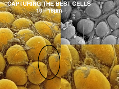
If you answered bone marrow, you are wrong. The answer is fat or adipose tissue which has 500 to 1,000 times the concentration of stem cells compared to bone marrow. Adipose-derived regenerative cells are today being used in clinical trials here in North America. Worldwide more than 10,000 patients have undergone regenerative medical treatment using their own MSCs. Here in Canada to date, there have been no clinical trials using MSCs. Canada’s only trials have used embryonic stem cells.
What is wrong with just doing trials with embryonic stem cells?
Embryonic stem cells, as promising as they have been, must be cultured in laboratories to grow a uniform population of sufficient volume for transplantation. What guarantees their genetic stability? The laboratory protocols covering generation and maintenance of cell populations is critical to ensuring that all embryonic stem cells function normally after transplantation without the potential for a mutation occurring in one or more of the cells. A mutation could lead to unintended consequences such as tumors with some potentially malignant.
Embryonic stem cells, when used in treatment, can be derived from the patient but the process to create them involves extracting pluripotent adult stem cells and reverting them to their embryonic state before culturing them to grow a population. Embryonic stem cells can also originate from a third party’s adult stem cells which through reversion can then become useful for regenerative therapies.
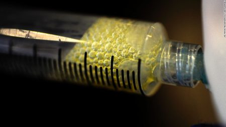
MSCs used in regenerative medicine procedures don’t require the complexity that is characteristic in using embryonic stem cells for therapy. They can be harvested from adipose tissue, our body fat, in large volumes, sufficient for therapeutic purposes and can be done with one particular technology (you’ll have to read Part 4 to find out which) in a single minimally invasive treatment process, done under local anesthetic, that involves three steps.
So what makes the transplanted MSCs differentiate?
With all cells sharing the same DNA it seems locality determines the type of differentiation that follows. So if you inject MSCs into a knee joint the MSCs will differentiate into chondrocytes. Put them into a liver and they will differentiate into the tissues found in that organ. Put them into a heart, or muscle and voila you will see differentiation produce the desired tissue.
How long does it take before results occur?
Differentiation begins almost immediately as cells replicate. But how does that translate in terms of time before the patient sees improvements? Depending on the medical issue a patient may begin to see improvements within a few days although with some conditions it may take up to six months for the regenerative treatment to work largely dependent on tissue type and normal replication rates through cell division.
In Part 4 of this series on regenerative medicine, I will introduce a technology that may prove to be a game-changer for regenerative medicine. Today, it is being used in a series of clinical trials around the world and its inventors are based in Richmond, British Columbia.
In this posting, I will talk about what makes stem cells tick? How do they go from being unspecialized cells to become specialized?
As scientists have learned more about stem cells they have also found out that they can be found in every tissue type in our bodies. In the past, they were overlooked because they would show up when looking at a slide of muscle tissue, bone, or lymph nodes, as small and innocuous cells with no known purpose. With hundreds of different cell types performing totally different body functions, cells that didn’t seem to do anything just didn’t ring any bells.
Our first discovery of stem cells came from studying embryos. Embryos begin as a ball of undifferentiated cells, the product of the initial division of a fertilized egg. But early on these undifferentiated cells take on specific tasks and eventually differentiate into hundreds of cell types. From first identifying the differentiating capability of embryonic stem cells, science soon moved on to discover stem cells elsewhere: in amniotic fluid, umbilical cord blood, and bone marrow.
One of the differentiated cells that come from stem cell precursors forms connective tissue. It is known as a stromal cell and is found in bone marrow, lymph nodes, ovaries, uterus, and prostate gland. Stromal cells come in two types: fibroblasts and pericytes.
Fibroblasts form connective tissue. They synthesize collagen which is responsible for providing the framework for various body tissue types.
Pericytes are the cells that help to form the superstructure for blood and lymph vessels.
Other types of body cells include a group of four with a common stem cell origin. These are osteoblasts which make up our bones, chondrocytes, our cartilage, myocytes, our muscles, and adipocytes, our body fat. Their common stem cell is referred to as mesenchymal (see illustration below).
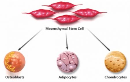
Mesenchymal stem cells (MSCs) are one of three types of stem cells responsible for building all the pieces that make us. MSC is to connective tissue as hematopoietic is to red and white blood cells, and platelets and endothelial is to blood and lymph vessels. The process that activates differentiation makes all of these three adult categories of stem cells produce differentiated organelles of various shapes, size, and featuring very different metabolic functions.
So what makes differentiation happen?
DNA, the essential molecule of life contains a blueprint of who we are going to be. It is found in genes on chromosomes forming the nucleus within our body’s cells. The DNA, RNA, and proteins within the cell all work in partnership. The DNA in the genes contains the bodybuilding instructions. The RNA and specific proteins execute the plan.
All this we understand. But we still don’t know the trigger that induces differentiation.
We can see it happen when we harvest stem cells and then inject them into an area in our body where they immediately begin to differentiate into the type of cells found there.
This is the state of regenerative medicine today.
Having said that we’ll conclude Part 2. In our next posting in this series, I will delve more deeply into the use of mesenchymal stem cells in therapeutic application. I’ll describe how these cells are harvested and the current technologies available to us. And I’ll also talk about current clinical trials where mesenchymal stem cells are being used for therapeutic regenerative medical treatment.
]]>

