To get the exact shape right the researchers used bright-field microscopy combined with nonlinear microscopy to create exact images of the targeted area within a simulated body. The iPSCs were encapsulated within a number of different bioinks to test the efficacy of each and determine which was most suitable for cartilage regeneration. After three weeks chondrocyte growth was indicated. The researchers noted that increasing the iPSC volume would likely speed up the proliferation of chondrocytes and the restoration of the body’s natural cartilage.
iPSCs don’t necessarily form chondrocytes. Being undifferentiated stem cells means that they could turn into almost any body tissue. So one of the issues the researchers had to address was ensuring that existing chondrocytes were at the locality where the bioink was introduced. That stimulated the iPSCs to turn into chondrocytes.
The researchers acknowledge that we still have a considerable way to go before we get to the point where it will be routine not to have knee or hip replacement surgery, but rather infuse stem cells into an affected joint to regrow lost cartilage to meet exact physical requirements. For me, the advancement in this technology cannot come soon enough to help me regenerate my left knee, and maybe even give my right one some fine tuning.
This latest research is being done at the Chalmers University of Technology, Gothenburg, Sweden, the Department of Clinical Chemistry and Transfusion Medicine, University of Gothenburg, and the Kungsbacka Hospital, in Kungsbacka, Sweden.

]]>
There have been many attempts over the years to create synthetic blood, a product that mimics blood’s essential purpose as the transporter of of oxygen and other nutrients throughout the body. But in every case no equivalent to natural blood has emerged.
Making blood from stem cells, however, would be a game changer. The goal would be to create the equivalent blood type, group O, rhesus negative. This is a type of blood that we define as the universal donor, a product suitable for all transfusion recipients. You see my blood can only be given to those who are also A Rh+ or AB Rh+. There are a lot of people on this planet with A Rh+ blood so in donating I can help them. There are very few with AB Rh+ and even so I can help them as well. But if all of this sounds a bit confusing here is quick blood type primer.
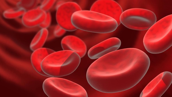
We have known since the early 20th century that not all blood is the same. That’s because the red blood cells each of us has contain different types of antigens. Antigens are sugars and proteins that can be found adhering to the surface of blood cells. Mismatched transfusions can trigger an immune system response. That’s why all who receive blood need to have their type identified before suitable donor blood can be dispensed. Our antigens are determined by our genetic makeup, our DNA. If our genes are not encoded for a particular sugar or protein then that antigen is absent from our blood cells. The absence or presence of these antigens determines the type of blood we have.
The identifying letters, “A,” “B,” and “O,” used here in North America and throughout much of the rest of the world (Russia and Eastern Europe use a numbering system) define the primary blood types. A secondary typing is known as “Rh.” And there are even more which we call subtypes, some of which are very rare. These are designated by the addition of a number beside the letter. So if you have your blood typed or if you choose to donate blood, you will be told that your blood is one of the following: A Rh+, A Rh-, B Rh+, B Rh-, AB Rh+, AB Rh-, O Rh+, O Rh-. Further testing may yield a sub-type such as A1, A1B, A2, A2B, and B1. These are among the 200 blood groups that modern science has identified.
Remember at the beginning of this posting I said that your blood type can tell you a bit about your origins. Here is a bit of blood geography lesson.
“A” blood type populations tend to be found in Central and Eastern European populations with one exception. The Blackfoot tribe in the United States happens to be predominantly type “A.”
“B” blood type populations are most commonly found in Asia, particularly China and India.
“AB” types are least common and found mostly in Japan, Korea and China.
“O” types, the most common are found in African, Hispanic and North American tribal populations.
The secondary blood typing “Rh,” which stands for rhesus factor, was first discovered in 1940 while testing inter-species blood transfusions between rhesus monkeys and rabbits., Rh+ is far more common than Rh-. Caucasian populations (European) tend to have the highest incidence of Rh- blood while Asian, Hispanic, Black, and North American aboriginal populations are predominantly Rh+.
Why is “O Rh-” considered the universal donor. Because both the “O” and the “Rh-” stand for the absence of the antigen proteins and sugars that cause immune responses.
Mismatching blood types has always had deadly consequences. Today such incidents are fairly rare. For example, in 2008 in the United States, 24 million transfusions were performed with 54 transfusion-related fatalities. In 2011 the number of transfusion deaths was 69. Before the turn of the 20th century mismatched blood deaths were common.
The goal of researchers in the United Kingdom, who have been working on producing blood from stem cells are trying to create a match to “O Rh-” for obvious reasons. This type of blood could be used by anyone. The researchers have chosen to work with embryonic and induced pluripotent stem cells (iPSCs). Starting with the embryonic type they can produce limited quantities of universal donor blood. But using iPSCs gives them access to much greater production volume. So the plan is to develop universal donor blood eventually from iPSCs. After four years the results are showing considerable promise with the first trials which they call “in-man,” scheduled for late 2016. “In-man” means exactly what it says. These trials will involve transfusing blood made from stem cells into human recipients.
One of the researchers in the program is Marc Turner, medical director of the Scottish National Blood Transfusion Service, describes the challenges and goal that lie ahead for the team. “Producing a cellular therapy which is of the scale, quality and safety required for human clinical trials is a very significant challenge, but if we can achieve success with this first in-man clinical study it will be an important step forward to enable populations all over the world to benefit from blood transfusions.”
In the meantime, I’ll still be donating blood every 56 days or so.


The research done by the Institute has created what are called multipotent stem cells. They cannot give rise to any cell type, merely a select subset. In this case skin cells have been able to create neural tissue. Schöler calls the process interconversion. It involves using a specific protein called BRN4. BRN stands for Brain. The protein is found in neural stem cells. Skin cells exposed to the protein become highly susceptible to conversion into neuronal somatic stem cells without reverting to the pluripotent state. When bathed in growth factors to increase cell division the skin cells’ interconversion speeds up with the cells losing their molecular memory. Within a few cell division cycles the conversion completes creating neuronal somatic stem cells that appear indistinguishable from normal stem cells.
Multipotency means stem cells like these can be used to regenerate damaged or diseased tissue. The next step is to duplicate the experiment using human skin cells and then track the stability of the cell line over time to ensure that it can become a safe source for cell and tissue regeneration.
]]>A Quick Primer on Heart Disease
Humans get two types of heart disease: congenital (that is born with) and acquired (disease that develops over time). Congenital tends to be structural. It can express itself in abnormal anatomy or physiology. Acquired begins with normal anatomy but physiology changes as the heart becomes diseased. Both can lead to heart failure which you can live with for a very long time or heart attacks which can lead to death.
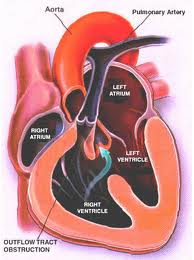
Structural defects to the heart and blood vessels in congenital heart disease can be repaired surgically, missing valves implanted and blood flow restructured using human grafts and artificial materials. Drugs can help regulate heart rhythm or improve the pumping efficiency. Pacemakers can help overcome structural problems that impact the electrical system that regulates heart rhythm.
Acquired, which represents the bulk of what we normally think of as heart disease, can also benefit from surgical intervention, drugs, pacemakers and other devices.
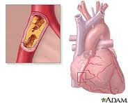
What causes heart disease? Lifestyle and inherited genes are the principal cause with 30 to 60% of the risk associated with the latter when it comes to acquired, rather than the former. Recent international studies identified 17 new genetic variants linked to increased heart disease risk. Five of these were associated with genetic regions that govern lipid metabolism processes, genetic controllers that impact the build up of fatty deposits in arteries. Ten others were in genes involved with other diseases and traits such as autoimmune diseases like Celiac and Type 1 Diabetes, cerebral and abdominal aneurysms, and lung cancer. What this means is there is no single genetic “smoking gun” that defines whether a person is likely to get heart disease in his or her lifetime. Instead we have a complex of genetic markers.
New Therapies for Repairing Bum Hearts and Blood Vessels
Stem Cells
In past blogs we have talked about stem cell therapy. Because stem cells have the potential to develop into a range of tissue types we can use them to regenerate and replace diseased tissue.
Cellular Dynamics International (CDI), is an American company located in Madison, Wisconsin, that is experimenting with the reprogramming of adult somatic cells to make them revert to a pluripotent state so that they can grow into any cell type needed for doing repairs to the body. These are induced pluripotent stem cells or iPS. Since 2008 CDI has used iPS cells to manufacture cardiomyocytes (heart muscle cells), freezing and sending them to pharmaceutical companies doing research on new drugs to treat heart disease.
But iPS cells have a brighter future if we can work out some of the bugs. Those bugs include faint traces of chemical residue from the parent cells used to create the reverted cells. Those residues can lead to mutations and as we have already written in previous blogs, mutations can lead to runaway cancer cell proliferation.
You can create iPS cells from almost any human tissue source. If a patient with a damaged heart provides a tissue sample, the cells from the sample can be induced to revert to iPS cells. They then can be transplanted back into the patient at the location requiring treatment. iPS cells from the donor are not rejected because they are from the donor who is also the recipient. The iPS cells in their reverted state can take on the characteristics of those cells that are in proximity to them and start to divide. iPS cells in the heart can generate heart muscle. iPS cells in the spine can generate nerve cells. CDI charges pharmaceutical companies $1,500 for 1.5 million iPS cells. That’s not cheap but the potential for iPS as an alternative therapy to transplants makes this a desirable area of ongoing research.
In research being conducted in Bristol, England and Glasgow, Scotland, Doctor Costanza Emanueli, along with a team of scientists are studying a variety of approaches to treating heart attack victims including using embryonic stem cells to create blood vessel cells to inject into patients. The method of delivery is a skin patch.
Gene Therapy
Researchers are looking at ways to tweak DNA to repair injured hearts. This involves manipulating a variety of genes that impact muscle and blood vessel growth.
Dr. Emanueli’s team, mentioned in the previous section, is studying boosting levels of nerve growth factor (NGF) to improve heart attack survival rates. In studies with mice the death rate from heart attacks was reduced by half using NGF. The gene responsible for NGF was attached to a specially engineered virus and delivered into the hearts of mice. Mice that received the NGF gene showed much improved circulation and heart function. In previous studies NGF was known to regrow nerves but in this case it encouraged new blood vessels to grow inside the injured heart muscle.
In another study conducted at Harvard’s Beth Israel Deaconess Medical Center, the gene C/EPB-beta spurred the growth of cardiomyocytes. This study showed that a genetic trigger responding to physical exercise turned on a molecular pathway to get cells to start dividing. For a very long time cardiologists could not find evidence of cell division in adult heart cells. Hearts under stress or subject to exercise would swell but cell proliferation in these enlarged hearts was poorly understood.
Heart muscle enlargement, called hypertrophy, can be associated with disease or with what is referred to as “athlete’s heart.” In the study cardiomyocyte growth was attributed to molecular events rather than factors such as high blood pressure.
Reprogramming Heart Cells After a Heart Attack
Heart attacks in the past have caused irreparable damage to heart muscles. What causes them? The usual suspects are accumulations of deposits on the walls of arteries that supply the heart with blood. When an artery is partly to fully blocked oxygen and nutrients are cut off to the area next to the blockage. This kills the muscle near the blockage affecting the heart’s ability to beat. But what if the section of damaged heart muscle can regrow?
Professor Paul Riley, of Oxford University’s Department of Physiology, Anatomy and Genetics, and his team of researchers are looking at a layer of cells known as the epicardium. The epicardium is the inner wall of the sac of tissue known as the pericardium that surrounds the heart. The embryonic epicardium contains many types of cells used by the heart and circulatory system including blood vessels and heart muscle. But after birth the epicardium gets quiet and stops generating these tissues. Dr. Riley and his team looked at ways to switch the epicardium cell generation back on. In mice studies they achieved just that by administering doses of Thymosin Beta 4, a natural occurring protein, to stimulate the embryonic genes in the epicardium to create cardiomyocytes. The researchers found that administering Thymosin Beta 4 before a heart attack proved to stimulate faster regrowth of heart muscle tissue.
The team continues to hunt for other naturally occurring molecules that can stimulate heart muscle and blood vessel growth. Along with drug and interventional therapies to clear arterial blockages, administering these proteins pre-emptively could create a bank of healthy new heart muscle to speed recovery should an attack occur.
Stem Cells
These life-changing undifferentiated cells are generated and harvested from 3 sources.
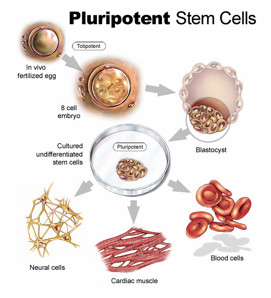
A human embryo in its earliest stages is called a blastocyst. It consists of two types of cells, an outer wall that becomes placental tissue and an inner wall of cells that eventually differentiate into all the tissues of the body. It is these latter undifferentiated cells that are potential tools for fighting disease and healing damaged tissue. This is a new field of reparative medicine done at the cellular level.
Stem cells can be reprogrammed. They have unique regenerative powers. Studying their growth is helping scientists to understand what causes birth defects. Currently researchers study stem cells for their potential to deal with a range of human diseases and problems including: stroke and traumatic brain injury, Alzheimer’s, Parkinson’s, blindness, deafness, traumatic wounds, ALS (amyotrophic lateral sclerosis), muscular dystrophy, diabetes, Crohns, cirrhosis of the liver, spinal cord injury, rheumatoid and osteoarthritis, heart disease, heart lesion repair after a heart attack, and many cancers. Researchers are even using stem cells to design and improve pharmaceuticals to target specific diseases and medical conditions.
Embryonic Stem Cells
We have experimented with embryonic stem cells since 1981 when scientists derived stem cells from the embryos of mice. By 1998 we discovered how to do this with human embryos. Only embryos discarded from in vitro fertilization procedures were used but nonetheless controversy soon erupted from religious groups that saw using these embryos as the equivalent of murder.
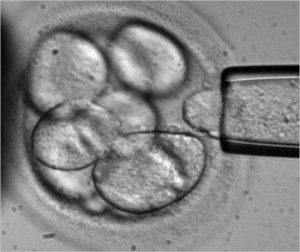
Embryonic stem cells can be cultured in a laboratory to divide while retaining their undifferentiated state. Called Pluripotent embryonic stem cells, they form stem cell lines which can be frozen and sent to laboratories and medical facilities for further research and testing.
In studying these cells researchers learn about human development and the complex processes involved when undifferentiated cells begin to differentiate to form various body tissues. This will help in understanding the mechanism that can lead to abnormal cell division and differentiation resulting in cancer and birth defects.
Somatic or Adult Stem Cells
Less controversial than embryonic, somatic (meaning cells not harvested from embryos) stem cells consist of undifferentiated cells found within body tissue that mostly contains differentiated cells. Somatic stem cells exist in organs and tissue in a caretaker role, providing maintenance and repair. We have been working with a form of somatic stem cells for some time. Probably the most well-known use is the procedure known as bone marrow transplantation used to treat leukemia, where hematopoietic stem cells are harvested from a donor and transplanted into a recipient.
Somatic stem cells have already been used to show their potential to create an endless supply of blood for transfusion allowing patients to bank cells that can be generated as needed eliminating the need for blood donors.
As researchers discover somatic stem cells in many more organs and body tissue their usefulness for other types of transplants may lead to cures for Parkinson’s, Alzheimer’s, heart disease, and many other intractable medical conditions.
Induced Pluripotent Stem Cells
Known in short form as iPSCs, these are adult tissue and organ cells that have been genetically reprogrammed to assume an undifferentiated embryonic stem-cell state. First “invented” and identified through the genetic manipulation of mouse cells in 2006, by 2007 human iPSCs had been harvested and cultured in laboratories.
Currently iPSCs are used for drug testing and modeling of diseases. Genetically altering an adult cell is no simple task. Using viruses as delivery agents, altered genetic materials get introduced into the cell. Unfortunately in some animal studies the viruses when introduced have caused cancer. We are not, therefore, ready to deploy iPSCs for therapeutic purposes in human studies. Researchers are exploring non-viral methods of delivering new instructions. Once they have mastered this the potential for therapies is enormous.
iPSCs can overcome tissue rejection. They can be used to repair and grow healthy tissue. This is the science of cell-based therapy and has the potential of replacing organ and tissue transplants. iPSCs can be created from a human source, cultured and directed to differentiate into specific cell types to treat diseases such as diabetes creating healthy insulin-generating cells in the pancreas, or repairing brains suffering from Alzheimer’s and Parkinson’s, or grow new heart muscle and blood vessels to cure heart disease, or repair immune systems that lead to osteo and rheumatoid arthritis. iPSCs have the potential to repair traumatic injuries such as spinal cord injury, or stroke, and regrow healthy skin to repair burns.
Stem Cell Therapy in the Medical Mainstream
How far are we away from seeing stem cell therapy in the medical mainstream? Today stem cells are being used to treat a small number of diseases and largely in clinical trial only. The most common use is blood stem cell transplantation for treating cancer. The PISCES study (Pilot Investigation of Stem Cells in Stroke), a fully regulated clinical trial of neural stem cell therapy for stroke patients started in 2010 in the U.K. In the United States clinical trials have started using embryonic stem cells differentiated into retinal cells to treat macular degeneration.
Soon these studies will be joined by many more. But first cell-based therapies need to move out of research laboratories. into many more clinical trials and then produce documented and consistent results to show that manipulated stem cells can be differentiated for transplantation consistently. That means creating enough cell volume before differentiation and controlling differentiation to create desired tissue types that match individual patients. That means ensuring that the tissues created from stem cells function the way they are supposed to for a lifetime and that the human host doesn’t experience rejection or develop any diseases resulting from transplanted cells.
We should be there if not within this decade then by 2030.
]]>


