COVID-19 began its assault on us as a respiratory infection causing severe damage to lung tissue. The medical establishment applied the same technology and treatments as it had done in the past for severe pneumonia and other deadly lung infections. Detection of the virus was done by taking nose swabs.
When I got COVID-19 in April 2020, the tests didn’t detect the presence of the virus. I didn’t present initially with lung issues. It was several weeks before I began coughing, a common sign of the infection. Instead, my voice changed, not for tracheal or bronchial reasons, but because my heart had enlarged and was pressing on my larynx.
An emergency doctor who saw me was the first to describe what I was experiencing. COVID-19 had infected my heart muscle and caused the left upper chamber to swell. She said that they were seeing an increase in symptoms similar to mine in the emergency department which they were presuming were related to COVID-19 exposure.
What I thought was indigestion when I felt changes in my chest turned out to be arrhythmias. The electrical conduction pathways of my heart were being altered by the virus infecting the contractile muscle cells responsible for pumping blood through the circulatory system. An electrocardiogram (ECG) showed I was in atrial fibrillation with my heartbeat jumping all over the place. Wearing a portable ECG, a Holter Monitor, for two weeks showed I was experiencing episodes of tachycardia (racing heart), bradycardia (slower than normal heart) and occasional moments when the muscle stopped beating lasting up to five seconds. The latter happened when I was sleeping which was a frightening thing to learn. COVID-19 could have killed me in my sleep. Three years later, having had two procedures and taking blood thinners, and anti-arrhythmic twice daily, my heart muscle is back to its prior calm self.
A study published earlier this year in Nature Cardiovascular Research that from March 2020 to March 2022, the United States saw 90,160 excess cardiovascular deaths higher than expected. The spikes in heart-related mortality appeared from March to June 2020 during the first wave of COVID-19, and from June to November 2021, a second noted COVID-19 death peak. The little over 90,000 represented a small percentage when compared to the global death count which surpassed 18.2 million during the first two years of the pandemic.
The majority of these excess deaths happened in people 65 years old or older. This is consistent with population health studies that show those of that age or older or more likely to experience cardiovascular events. A significant number of deaths associated with the heart found the biggest spike, however, in young adults between the ages of 25 and 44. These included higher incidents of heart attacks, strokes and arrhythmias associated with COVID-19 with occurrences happening up to a year after the initial infection.
A 2022 Lancet study of 42 million U.S. medical records covering from January 2019 to March 2022 ended up recruiting over 4 million participants who had been tested for COVID-19. The study published in eClinicalMedicine revealed a much higher risk of heart-related diseases occurring in the year after having tested positive for COVID-19. Higher incidents of stroke, heart attacks, arrhythmias, myocarditis, ischemic cardiomyopathy, heart failure, hypertension and pulmonary embolisms appeared in these medical records.
Dr. Helene Glassberg, a cardiologist at the University of Pennsylvania Perelman School of Medicine, is quoted in a New York Times, September 7, 2023, article, stating “Early on, we saw the impact on the cardiovascular system during Covid illness…But now we’re seeing the long-term consequences in people who’ve had prior Covid.” Dr. Glassberg was not a participant in the Lancet study but concurs with what the data shows. Although there have been rare incidents of myocarditis occurring from vaccinated patients, something that vaccine skeptics like to point out, the risk of getting myocarditis from the virus is 28 times greater.
A Children’s Hospital of Philadelphia (CHOP) led research study published in Science Translational Medicine in August 2023 points to COVID-19-induced mitochondrial genetic changes as the possible agent leading to heart and other organ damage from the virus. The mitochondrion is an organelle found in every human cell. It has separate DNA and genes from the cell’s nuclear DNA.
Evidence shows the proteins found in COVID-19 have a fondness for mitochondrion. They bind with the organelle’s proteins to suppress mitochondrial gene expression. So, although the initial access for COVID-19 may be through our lungs, the damage to its mitochondria is temporary. Lung cells recover. That is not the case, however, in other organs, particularly in the heart, where damage is long-term.
States Douglas C. Wallace, Director of the Center for Mitochondrial and Epigenomic Medicine (CMEM) at CHOP, “We need to stop looking at COVID-19 as strictly an upper respiratory disease and start viewing it as a systemic disorder that impacts multiple organs. The continued dysfunction we observed in organs other than the lungs suggests that mitochondrial dysfunction could be causing long-term damage to the internal organs of these patients.”
Previous studies may have identified a mitochondrial function regulator that could lead to a new therapy for those suffering from Long COVID. Called microRNA 2392 (miR-2392), it could impede viral replication to stop long-term complications from COVID-19.
]]>About fifteen years ago bloodwork ordered by my family doctor indicated my cholesterol levels were a little above high normal. My doctor prescribed a statin which I continue to take a minimal dose each day. It took awhile for me to adjust to a side effect – lower back pain, but by adding CoQ10 to my daily intake I have had no problems.
High cholesterol levels in the blood are a potential heart disease threat because an excess can cause fatty deposits to grow on the walls of blood vessels which over time can lead to blockages, a stroke, or a heart attack.
High cholesterol can be an inherited condition. It can also occur from an unhealthy diet, other medications, or a lack of exercise. The prevalence of high cholesterol in North American populations has had some in the medical profession suggesting that statins should be added to the drinking water, just like fluoride used to prevent tooth decay.
Cholesterol in combination with proteins expressed through genetic instructions produces a substance called lipoproteins. There are two versions: low-density lipoprotein (LDL) and high-density lipoprotein (HDL). It is LDL that causes fatty deposits. HDL is seen as good because it removes excess cholesterol from the bloodstream.
Taking statins targets LDL and in some studies is seen as increasing HDL levels in blood plasma. But it is still unclear whether statins impact HDL effectiveness.
But what if we didn’t need statins and could end high cholesterol by altering a specific gene associated with blood cholesterol levels? In Peter Diamandis’ latest musings he describes what appears to be a biomedical game changer. A therapy that permanently alters one gene responsible for a protein that produces LDL and HDL.
I have edited Peter’s work and hope you find what follows to be an interesting read.
Heart disease accounts for roughly 16% of total deaths globally each year. It costs the US economy roughly $219 billion annually. High levels of cholesterol in the bloodstream is one of the main causes. That’s why more than 200 million people worldwide regularly take statins, the leading medication for lowering cholesterol levels. These drugs have proved effective, but require consistent use and can result in side-effects.
Scientists have identified a link between the PCSK9 gene and a protein for which it is responsible that plays a role in regulating cholesterol levels in the blood.
Research at the University of Texas Southwestern Medical Center in the mid-2000s established the role of the PCSK9 gene in heart health. The researchers identified people with a genetic mutation that inhibited it from producing a protein had lower LDL levels in their bloodstreams.
The PCSK9 gene-produced protein was seen as preventing the recycling of receptors in the liver responsible for filtering LDL from the bloodstream. Where the PCSK9 gene produced high levels of the protein the overall number of receptors dropped and cholesterol levels rose. The impact of the amount of protein produced by the PCSK9 gene was associated with 15 to 28% reductions in LDL levels and a 47 to 88% lower risk of heart disease.
Finding a drug that could target the protein has proven challenging. Monoclonal antibodies that target PCSK9 called alirocumab and evolocumab have been produced to cut the risk of cardiovascular events by half. But these drugs are more expensive than statins.
The Texas Southwestern researchers thought that a more promising approach would be to tackle the errant gene rather than the protein it produces. From the patients they identified that had mutations and low LDL levels, it seemed possible to genetically replicate a resistance to heart disease by changing all PCSK9 genes to express less protein.
The tool of choice for this project was CRISPR. The emergence of the CRISPR-Cas9 system has revolutionized our ability to tweak genes. There are issues however that have limited its applicability in biomedicine. That’s because the Cas9 protein produced by CRISPR cuts both strands of DNA at a target site before relying on the cell’s internal repair system to stitch the DNA together. Experience has shown that the process isn’t always perfect leading to unintended edits that reduce efficacy and can potentially introduce harmful mutations.
An emerging approach known as base editing gets around the problem by using a modified Cas9 protein to break only one of the DNA strands. This is spliced to another protein to convert one DNA letter to another. The approach only makes single letter edits but is much more precise for use in medical applications. And by a remarkable stroke of luck, it turns out that the PCSK9 gene can be altered to deal with the errant protein through a simple one-letter edit. This is a big deal because diseases caused by a single-point mutation like this are relatively uncommon.
A company called Verve Therapeutics has designed a base editor to switch out a single letter in the stretch of DNA encoding for PCSK9 to essentially turn the gene off. Last year, they reported when they injected the therapy into the livers of monkeys it reduced their LDL levels by 60%.
The first human trial began this month with the first to receive the therapy a New Zealand volunteer. The plan is to treat 40 people who have an inherited condition called familial hypercholesterolemia (FH) which occurs in 1 in 250 and produces extremely high levels of LDL. Those with FH are at higher risk for heart disease. If successful the treatment can become a standard approach to dealing with FH and other high cholesterol risks. Stated Sekar Kathiresan, CEO of Verve, in a recent MIT Technology Review article, “If this works and is safe, this is the answer to heart attack—this is the cure.”
The therapy is a one-and-done treatment unlike being on statins or taking monoclonal antibodies. It will make a permanent change to liver cells and stop them from producing the PCSK9 protein. The edits will pass on to the next generation of cells when they divide. That means that even if the therapy is initially expensive, the lifetime cost should prove to be comparable if not lower than current alternatives.
The therapy is delivered to the liver cells in lipid nanoparticles, the same technology being used by mRNA Covid-19 vaccines. The pandemic has created manufacturing capacity for this type of therapeutic delivery which is helping to lower costs.
A successful clinical trial doesn’t lead right away to adoption throughout the medical profession. It is likely there will be many more trials, and a number of years before Verve has enough data to convince regulators to approve the treatment for run-of-the-mill high cholesterol. If it turns out to become the new treatment standard, however, it will be one of the most transformational innovations in human healthcare to be seen in decades.
Beyond the potential to cure heart disease, this news is an exciting milestone in the use of genetic engineering in clinical application. Current base editing technology is capable of tackling about 60% of single-point mutations associated with diseases and this could widen the scope for its use.
Beam Therapeutics is another company working with base editing technology that is about to start human clinical trials designed to treat the genetic blood disorder sickle-cell anemia. It is using base editors to make multiple single letter changes for more complex genetic repairs.
Then there is Wave Life Sciences that is experimenting with base editors to modify RNA rather than DNA to make it possible for transient changes to how genes are expressed rather than permanent alterations to the genome.
Most excitingly, the researcher who first developed base editing, David Liu from the Broad Institute of MIT and Harvard, has continued innovating. In 2019, he and his colleagues outlined a new approach called prime editing in the journal Nature, which dramatically increased the scope of potential edits. Base and prime editing are being used by the team at CureHeart to tackle cardiomyopathies. You can read what I recently wrote about their work on this blog site. Like base editing, prime only breaks a single DNA strand, but in addition makes edits to individual letters or entire stretches of genetic sequences. It is believed that up to 89% of mutations associated with diseases in humans can be treated this way.

In his mid-80s, my father-in-law (seen in the above picture at an earlier time in his life) suddenly found walks from his midtown home to his jewellery store in the heart of downtown Toronto, were suddenly getting much harder. He described what he was experiencing as walking wearing concrete shoes. He would become breathless at times. This was a man who cycled 20 kilometres almost every weekend. Referred to a cardiologist, he learned he had a genetic disorder, hypertrophic cardiomyopathy, and that its appearance in his 80s was indeed rare.
Hypertrophic cardiomyopathy is one of those heart ailments with a genetic link. It causes the heart muscle to thicken losing its flexibility as a pumping chamber. As the heart wall, particularly the septum which separates the left and right ventricles, thickens it begins to impede blood flow through the aortic valve to the rest of the body.
Although hypertrophic cardiomyopathy can happen at any age, it is rare to begin expressing symptoms in someone as late as it did with my father-in-law. Most of those who get it have a family history of the disease. The odds of inheriting the genes are 50/50. That’s why my wife is followed by a cardiologist regularly.
At the time my father-in-law was given his diagnosis he was offered two options. One involved an operation to ablate, that is burn away a part of the thickening walls, to restore flexibility. The second was a pacemaker. He chose the latter.
Cardiomyopathies occur in about 1 in every 250 people. In the U.S. approximately 1.5 million are affected. Many are unaware. It appears in all life stages. In addition to hypertrophic there are other cardiomyopathies. These include:
People with cardiomyopathy more often find their lives shortened although not in the case of my wife’s family. My father-in-law died at age 97. His father had it but lived to age 92. And his sister also was diagnosed with it and died at age 102. It looks like they had other genes that were working in their favour.
Cardiomyopathies are life-threatening. They cause arrhythmias and can lead to a stroke, heart failure or even a heart attack. When young people get diagnosed with hypertrophic cardiomyopathy the option today is a heart transplant or a very shortened lifespan. With arrhythmogenic right ventricular dysplasia in young people, defibrillators can be implanted to stop the arrhythmia and a heart transplant is the only other option.
CureHeart was formed by a group of researchers from a global community that includes the University of Oxford, Harvard, the Broad Institute, Duke, Share, Wave Life Sciences, Myokardia, and Cardiomyopathy UK, a heart muscle charity. Their work on gene therapy for cardiomyopathy recently won them the British Heart Foundation’s Big Beat Challenge, a contest that involved 75 entries from around the world competing for a £30 million grand prize.
The CureHeart team uses a technique called base and prime editing to suppress and eliminate faulty genes linked to inherited heart muscle diseases. They have identified specific genes that work alone or in combination with others responsible for genetically inherited heart conditions like cardiomyopathies. Mice studies are showing that in some cases a faulty gene can be repaired or switched off. But cardiomyopathies can involve a cascade of gene interactions following a specific sequence which means treating them in isolation isn’t sufficient. So CureHeart is developing a way to switch off faulty genes, edit and correct them, and do it in correct sequence.
The plan is to deliver a cure through a vaccine which can be administered like any other, a shot in the arm, or can be introduced through the chest wall directly into the heart muscle itself. The end product would be offered to family members with a history of genetically-associated cardiovascular disease.
As a parent of a child born with congenital heart disease, the CureHeart project represents an exciting development for coming generations. The team notes that what can work for cardiomyopathy could prove to be useful for curing other inheritable cardiovascular diseases. And it doesn’t have to stop at the heart considering the numerous other gene-associated diseases that humanity faces. If successful, vaccine-delivered cures like these could become standard treatment in reversing bad genes. This medical advance will be a remarkable achievement.
]]>The first involves the use of AI to identify areas within human organs that surgeons can safely dissect before operating on a patient. The second is the ability for AI to predict if memory issues in patients will develop into dementia within two years. And the third is the use of AI to analyze eye scans done during routine visits to an optician to identify patients at high risk for a heart attack.
In Toronto, Dr. Amin Madani, a general surgeon at the University Health Network, has developed a promising use for AI to improve surgical procedures and patient outcomes. Using deep learning, a subset of machine learning, Dr. Madani has trained an AI model by studying thousands of surgical procedure images involving dissection of livers, gallbladder, and other organs to identify safe and dangerous areas within organs’ anatomical structures to provide guidance during operations. The AI was able to score go and no-go zones for intervention with considerable accuracy.
In an article posted to CBC News in December of last year, a surgical instructor from Case Western Reserve in Cleveland, who collaborated with Dr. Madani is quoted stating, “The hope is to…bring a second pair of eyes into the operating room, in this case, machine eyes, to ensure the surgeon is seeing whey they think they’re seeing.”
How does this deep learning AI work? When a surgeon uses laparoscopic techniques to do gallbladder and other organ procedures. a camera gets inserted through the slit and projects images of the surgical field on a screen. What the AI does is project colours onto the images the surgeon sees. A green-coloured area of the surgical field is safe to cut. Coloured areas that are red are not.
The progress being made by Dr. Madani and his collaborators was first published in 2020 in the Annals of Surgery journal. The initial organ of choice has been the gallbladder since it is one of the most common operations and relatively easy to fully document with imaging from thousands of successful procedures. Based on the initial success the plans are to expand its use for other organs and a wider range of surgical interventions.
Machine learning (ML) is proving to be an effective AI tool in helping to identify false diagnoses of dementia from those who suffer from it in its most progressive form leading to an Alzheimer’s Disease diagnosis.
A study done at the University of Exeter in the United Kingdom shows that in looking at data from 15,307 individuals attending memory clinics, its AI tool with high accuracy predicted which of them would develop dementia within two years.
Clinical assessment tools exist for predicting dementia in patients over the longer term, but the challenge for clinicians working in this field is to assess patients who in the short term might be at high risk. Prior to the development of the ML tool, no clinical decision-making tool had previously been developed for identifying likely Alzheimer’s diagnoses within a window of one to two years.
How does this AI work? The ML tool studied the files of patients being followed between 2005 and 2015. All of the files the ML looked at were of patients who did not have dementia upon initial clinical examination. Many exhibited memory issues or had problems with other brain functions. But the ML tool was able to establish patterns within the patient record and identify the level of risk with much higher accuracy than human clinicians. It accurately diagnosed 92% of patients who developed Alzheimer’s within two years. It also accurately identified 80% of inconsistent diagnoses. In the study’s conclusions, it noted that the ML tool has the potential to reduce misdiagnoses by up to 84%.
The research lead who oversaw the study, Dr. David Llewellyn, at the University of Exeter, is quoted in an article appearing on the Biotechnology Community website, “We’re now able to teach computers to accurately predict who will go on to develop dementia within two years. We’re also excited to learn that our machine learning approach was able to identify patients who may have been misdiagnosed. This has the potential to reduce the guesswork in clinical practice and significantly improve the diagnostic pathway, helping families access the support they need as swiftly and as accurately as possible.”
A deep learning AI being developed at the University of Leeds in the United Kingdom has been trained to read retinal scans and identify patients who were likely to have a heart attack within a year. The study’s results appeared in the last week in the journal, Nature Machine Intelligence.
How does the AI tool work? Changes to blood vessels in the retina are indicators of vascular disease complications. This has been known by doctors for some time. But which patients go on to develop heart attacks within a short window of time remained an unknown. The deep learning AI analyzed retinal scans and cardiac scans from more than 5,000 patients. It was able to identify what changes it saw in the retina with changes in the heart. Once the patterning was learned the AI was able to estimate the pumping efficiency and size of the heart’s left ventricle which if enlarged is directly linked to increased risk of a heart disease. This information along with a review of the patients, age, and sex, led to a tool capable of accurately predicting 70 to 80% of the time if the patient was at short-term risk of a heart attack occurring within 12 months.
As a diagnostic tool, this allows doctors through routine and inexpensive eye screening examinations to intervene with early treatment to prevent cardiovascular disease leading to a major heart incident.
One of the researchers involved in the study, Professor Sven Plein at the university, is quoted in a press release stating, “The AI system is an excellent tool for unravelling the complex patterns that exist in nature, and that is what we have found – the intricate pattern of changes in the retina linked to changes in the heart.”
]]>Early into the pandemic, cases in Wuhan and at Massachusetts General Hospital observed the elevated troponin phenomenon in patients with prior cardiovascular conditions such as congestive heart failure, coronary artery disease, and hypertension, as well as in those without a previous history. of heart problems Soon other hospitals were reporting and documenting elevated troponin levels in COVID-19 cases causing myocarditis, an inflammation of the heart muscle, as well as arrhythmias, heart failure, and even cardiac arrest, and sudden death.
The phenomenon first observed in acute COVID-19 infections also started showing up in cases with milder symptoms, and in asymptomatic patients. In a report published in JAMA in July 2020, 78 patients out of 100 who had recovered from the common symptoms associated with COVID-19 in magnetic resonance imaging (MRI) tests showed ongoing myocarditis. Of these patients, 60 had never had a previous history of cardiovascular disease. The July study included two-thirds of the 100 in total who never required hospitalization. Many of those studied were asymptomatic.
Later in the year, a September 2020 study looking at a small sampling involving 26 athletes from The Ohio State University who had been infected with COVID-19, showed scarring of the heart in 8 and prolonged myocarditis in 4. In this study, none of the athletes had shown elevated troponin levels. Scans were done 11 to 53 days after a two-week quarantine.
A December case published in JACC, the Journal of the American College of Cardiology Foundation, reported elevated troponin levels and heart failure symptoms in a 2-month-old infant infected with COVID-19.
What has concerned doctors is that COVID-19 patients may have long-term heart problems that last well beyond the end of the viral infection. Another issue of concern is that the myocarditis being observed in many cases is atypical. In other words, not the usual type of myocarditis seen from other medical conditions.
Some physicians speculate that the elevated troponin levels, and subsequent myocarditis may not just be from COVID-19, but potentially may come from other viruses that opportunistically attack the body in its weakened state.
So elevated troponin levels more often than not are linked to COVID-19 and abnormal imaging findings of patients’ hearts and blood vessels. This is not a universal rule. But it is a pretty good indicator in the majority of researched cases.
The concern by doctors now is just how long these conditions persist after the infection has passed. A new clinical trial has been proposed to follow 900 COVID-recovered patients for a period of three years to determine just how long cardiac symptoms and potential toxicity last.
]]>
Ding is using CRISPR/Cas9 to target genes associated with these diseases. She states “heart disease and high cholesterol are associated with certain gene mutations. We introduce these gene mutations into human pluripotent stem cells….” By doing this she and her team of researchers study the mutations and find ways to treat them in a petri dish. She has been doing it successfully with mouse genes which brings the question up, when will we be able to do it with humans?
Ding believes we are still a few years away from trying this on humans largely because of potential unintended consequences should a therapy impact more than the disease it targets. But the advantages of editing an existing genome are self-evident. It’s a permanent fix.
What Ding is doing is far from eugenics and designer babies although one could see the technology being used in this way. She restricts her research to non-reproductive tissues, liver and muscle cells. Altering the genome of a parent to correct a congenital disease (congenital means born with) does not protect future generations from inheriting the disease since sperm and egg cells are not within the realm of Ding’s research.
But that is coming as embryologists look at ways of intervening early in the development process to alter an inheritable genetic defect to ensure a healthy baby is born.
Ding believes we can eliminate almost all genetic diseases with this technology and line of research. And ultimately she believes all disease can be eliminated through gene editing.
You can read an interview given by Ding at the World Economic Forum at the link provided here.

]]>
The first clinical trial is underway headed up by Dr. Amit Patel, Director of Clinical Regenerative Medicine and Tissue Engineering, and his associate Dr. Craig Selzman. For the 6 million humans living with heart failure this new therapy may eliminate the need for transplants and more invasive surgical procedures.
The procedure which goes by the name of Athena, is done on an outpatient basis and involves the use of a cardiac catheter which delivers an infusion of human DNA, called SDF-1, directly to the site within the heart or a coronary artery where the problem exists. SDF-1 stands for stromal cell-derived factor-1, a chemokine (a signaling protein that cells secrete) that has significant therapeutic value in tissue engineering. When a body sustains an injury the damaged cells emit the chemokine to attract the mechanisms within the body to do self-repair. The Utah team in injecting SDF-1 to a heart-damaged area are inducing within the body a super-response acting as a homing beacon for the body’s own restorative mechanisms.
The first successful procedure, performed on Ernie Lively (seen in inset image below), an actor known for his appearances in several films, occurred on November 7. Lively is recovering very well according to the Utah team who have 72 other patients in the wings waiting for the procedure.
You can watch the procedure on YouTube. It doesn’t look like much but it may usher in a new age for treatment of many types of heart disease.

But donor demand far exceeds supply with only 4,000 hearts available for more than 100,000 on wait lists. That’s why artificial hearts, mechanical devices that mimic the heart’s beat, are being developed to act as bridging devices to extend a patient’s life until a heart becomes available. And in some cases manufacturers are hoping to develop devices that can replace the natural heart entirely.
One of these companies is SynCardia Systems, located in Tucson, Arizona. It has built an artificial heart that today is implanted in more than 1,100 human patients accounting for more than 300 patient-years of use. The SynCardia artificial heart combines an implant, seen below, with an external control system that the patient wears. Tubes are inserted through the skin. The tubes pump air into two small balloons which then push blood through the implant to the circulatory system. SynCardia is not meant to be a permanent replacement for a real heart. But it does give patients some freedom from hospitalization while waiting for a transplant. In 2012 the Federal Drug Administration in the United States approved SynCardia as a bridging device. The company’s ultimate goal is to have future version of the device approved for permanent use in cases where patients are not transplant eligible. Currently the biggest drawback to the SynCardia implant is the 6 kilogram (13.5 pound) portable, wearable power supply that constantly needs recharging every 4 to 6 hours.
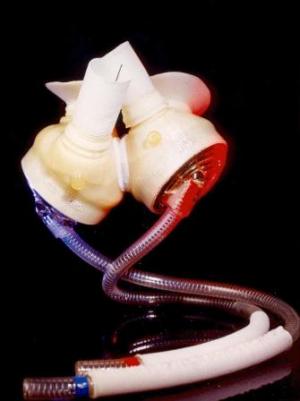
Carmat, a Paris-based company, hopes to improve on all aspects of the SynCardia solution. Its artificial heart combines biology with mechanical components. A membrane made from a cow’s pericardium (the tissue that surrounds the heart) separates the moving parts from the blood system minimizing the need for the patient to be on anti-coagulants. The Carmat is truly a biomimetic device. It looks a little bit like a heart (see the picture below) and its valves are made from biological tissue. In addition it uses 12-hour rechargeable lithium-ion batteries weighing 3 kilograms (6.6 pounds) to power its two artificial pumping chambers, each filled with hydraulic fluid, not air. This means no large tubal inserts through the skin into the body. It is the movement of the fluid between the pumping chambers that mimics the natural heart while never interacting with blood. And Carmat plans to replace its current battery pack with a portable fuel cell making the system the first to use such a power supply in a medical device.
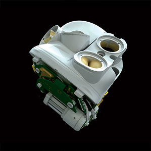
But SynCardia and Carmat are not alone. Last year I wrote about the HeartMate II, a left-ventricle assist device that has been implanted in thousands around the world. And an Australian biomedical engineer, Daniel Timms, has teemed up with the doctors at the Texas Heart Institute to develop an artificial heart that doesn’t beat. Called the BiVACOR artificial heart, it uses a magnetic field, centrifugal force and a single moving part, a spinning disc, to push blood through the body. The prototype can be seen in the picture below. It is hoped by the medical team at the Texas Heart Institute that they can begin human medical trials in a few years.

]]>
A Quick Primer on Heart Disease
Humans get two types of heart disease: congenital (that is born with) and acquired (disease that develops over time). Congenital tends to be structural. It can express itself in abnormal anatomy or physiology. Acquired begins with normal anatomy but physiology changes as the heart becomes diseased. Both can lead to heart failure which you can live with for a very long time or heart attacks which can lead to death.
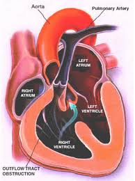
Structural defects to the heart and blood vessels in congenital heart disease can be repaired surgically, missing valves implanted and blood flow restructured using human grafts and artificial materials. Drugs can help regulate heart rhythm or improve the pumping efficiency. Pacemakers can help overcome structural problems that impact the electrical system that regulates heart rhythm.
Acquired, which represents the bulk of what we normally think of as heart disease, can also benefit from surgical intervention, drugs, pacemakers and other devices.
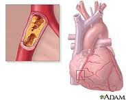
What causes heart disease? Lifestyle and inherited genes are the principal cause with 30 to 60% of the risk associated with the latter when it comes to acquired, rather than the former. Recent international studies identified 17 new genetic variants linked to increased heart disease risk. Five of these were associated with genetic regions that govern lipid metabolism processes, genetic controllers that impact the build up of fatty deposits in arteries. Ten others were in genes involved with other diseases and traits such as autoimmune diseases like Celiac and Type 1 Diabetes, cerebral and abdominal aneurysms, and lung cancer. What this means is there is no single genetic “smoking gun” that defines whether a person is likely to get heart disease in his or her lifetime. Instead we have a complex of genetic markers.
New Therapies for Repairing Bum Hearts and Blood Vessels
Stem Cells
In past blogs we have talked about stem cell therapy. Because stem cells have the potential to develop into a range of tissue types we can use them to regenerate and replace diseased tissue.
Cellular Dynamics International (CDI), is an American company located in Madison, Wisconsin, that is experimenting with the reprogramming of adult somatic cells to make them revert to a pluripotent state so that they can grow into any cell type needed for doing repairs to the body. These are induced pluripotent stem cells or iPS. Since 2008 CDI has used iPS cells to manufacture cardiomyocytes (heart muscle cells), freezing and sending them to pharmaceutical companies doing research on new drugs to treat heart disease.
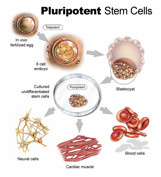
But iPS cells have a brighter future if we can work out some of the bugs. Those bugs include faint traces of chemical residue from the parent cells used to create the reverted cells. Those residues can lead to mutations and as we have already written in previous blogs, mutations can lead to runaway cancer cell proliferation.
You can create iPS cells from almost any human tissue source. If a patient with a damaged heart provides a tissue sample, the cells from the sample can be induced to revert to iPS cells. They then can be transplanted back into the patient at the location requiring treatment. iPS cells from the donor are not rejected because they are from the donor who is also the recipient. The iPS cells in their reverted state can take on the characteristics of those cells that are in proximity to them and start to divide. iPS cells in the heart can generate heart muscle. iPS cells in the spine can generate nerve cells. CDI charges pharmaceutical companies $1,500 for 1.5 million iPS cells. That’s not cheap but the potential for iPS as an alternative therapy to transplants makes this a desirable area of ongoing research.
In research being conducted in Bristol, England and Glasgow, Scotland, Doctor Costanza Emanueli, along with a team of scientists are studying a variety of approaches to treating heart attack victims including using embryonic stem cells to create blood vessel cells to inject into patients. The method of delivery is a skin patch.
Gene Therapy
Researchers are looking at ways to tweak DNA to repair injured hearts. This involves manipulating a variety of genes that impact muscle and blood vessel growth.
Dr. Emanueli’s team, mentioned in the previous section, is studying boosting levels of nerve growth factor (NGF) to improve heart attack survival rates. In studies with mice the death rate from heart attacks was reduced by half using NGF. The gene responsible for NGF was attached to a specially engineered virus and delivered into the hearts of mice. Mice that received the NGF gene showed much improved circulation and heart function. In previous studies NGF was known to regrow nerves but in this case it encouraged new blood vessels to grow inside the injured heart muscle.
In another study conducted at Harvard’s Beth Israel Deaconess Medical Center, the gene C/EPB-beta spurred the growth of cardiomyocytes. This study showed that a genetic trigger responding to physical exercise turned on a molecular pathway to get cells to start dividing. For a very long time cardiologists could not find evidence of cell division in adult heart cells. Hearts under stress or subject to exercise would swell but cell proliferation in these enlarged hearts was poorly understood.
Heart muscle enlargement, called hypertrophy, can be associated with disease or with what is referred to as “athlete’s heart.” In the study cardiomyocyte growth was attributed to molecular events rather than factors such as high blood pressure.
Reprogramming Heart Cells After a Heart Attack
Heart attacks in the past have caused irreparable damage to heart muscles. What causes them? The usual suspects are accumulations of deposits on the walls of arteries that supply the heart with blood. When an artery is partly to fully blocked oxygen and nutrients are cut off to the area next to the blockage. This kills the muscle near the blockage affecting the heart’s ability to beat. But what if the section of damaged heart muscle can regrow?
Professor Paul Riley, of Oxford University’s Department of Physiology, Anatomy and Genetics, and his team of researchers are looking at a layer of cells known as the epicardium. The epicardium is the inner wall of the sac of tissue known as the pericardium that surrounds the heart. The embryonic epicardium contains many types of cells used by the heart and circulatory system including blood vessels and heart muscle. But after birth the epicardium gets quiet and stops generating these tissues. Dr. Riley and his team looked at ways to switch the epicardium cell generation back on. In mice studies they achieved just that by administering doses of Thymosin Beta 4, a natural occurring protein, to stimulate the embryonic genes in the epicardium to create cardiomyocytes. The researchers found that administering Thymosin Beta 4 before a heart attack proved to stimulate faster regrowth of heart muscle tissue.
The team continues to hunt for other naturally occurring molecules that can stimulate heart muscle and blood vessel growth. Along with drug and interventional therapies to clear arterial blockages, administering these proteins pre-emptively could create a bank of healthy new heart muscle to speed recovery should an attack occur.

