Before I underwent knee replacement surgery three years ago last July, I was hellbent on waiting for a stem cell therapy alternative to regrow my arthritic joint. Having endured more than a decade of discomfort and a course of cortisone injections to keep the surgeons at bay, the advances in stem cell research had not yet reached a point where it could be considered a non-surgical alternative therapy.
There were several clinical trials. The closest still accepting volunteers was in Chicago. The only one in Toronto had reached its complement of candidates. Eventually, I was referred to an orthopaedic surgeon who explained to me why knee replacement using stem cells to regrow cartilage was still seeking answers for its most challenging limitation.
Mesenchymal stem cells harvested from a patient could be turned into chondrocytes, the name given to cartilage cells. Cartilage cells, however, need a matrix upon which to grow. That natural matrix is made of collagen, a structural protein our bodies produce that forms connective tissue. At the time of my referral, no stem cell treatment had yet to come up with a way to produce collagen and put it where it was needed to support growing cartilage.
The treatment of Liesel, the elderly gorilla, used mesenchymal stem cells harvested from a younger donor female gorilla who had undergone minor surgery in 2022. StemCellX, a company formed by leading stem cell scientists, worked with researchers from the University of Sheffield in the United Kingdom on Liesel’s treatment.
StemCellX is focused on cutting-edge research and stem cell treatments for veterinary medicine. It provided the harvested mesenchymal stem cells that were kept frozen until the procedure. There was no mention of concern about Liesel rejecting the stem cell infusion. That’s because mesenchymal cell transplants exhibit low rejection rates and tend to be well tolerated in transplantation. There was high confidence that Liesel would benefit since StemCellX has successfully treated dogs and horses with moderate to advanced osteoarthritis.
Several clinical trials using mesenchymal stem cell therapies for treating osteoarthritis have recently reported success. One from Brazil documents the use of bioscaffolds made up of collagen or hyaluronic acid applied to knee joints in place of natural collagen with intra-articular injections of mesenchymal stem cells to induce cartilage regeneration in osteoarthritic knee joints. The following illustration appears in the journal article that describes this clinical trial.

In another two-year case study published in the journal, Regenerative Medicine, 329 patients with knee osteoarthritis received mesenchymal stem cell therapy with significant improvements to the knee joint. In this clinical trial, the goal was to reduce or delay the need for total joint replacement.

In a University of California, Irvine (UC Irvine) clinical trial, laboratory-made neurons grown from embryonic stem cells have been injected into the brains of 12 patients with Parkinson’s. The clinical trial is ongoing but initial results are encouraging showing that the added neurons have formed synapses to link to the patients’ brain cells and produce dopamine.
Dopamine is a neurotransmitter used to send signals to neurons in the brain. Naturally produced by our bodies, too much dopamine causes aggressive behaviours in people Too little leads to diseases like Parkinson’s, restless leg syndrome and depression. Dopamine is often called the pleasure molecule because when produced it can induce those types of feelings. Getting the dopamine balance right, therefore, has turned out to be a way to treat Parkinson’s.
In the UC Irvine study, PET scans have been used to observe how the brain’s uptake of the neurons is succeeding. After a year, these laboratory-produced neurons appear to be surviving while Parkinson’s symptoms are shown to be lessening. The university’s ongoing research into Parkinson’s is not only focused on delaying the onset of the disease but also rebuilding the brains of patients suffering from it. The ongoing trial which started in May 2021 will conclude in 2024. At that time and based on reported success, the plans are to increase the number of Parkinson’s patients receiving neurons created from embryonic stem cells.
In the United States, embryonic stem cell research is considered highly controversial. Those opposed to a woman’s right to choose associate this type of research with abortion with the harvesting and use of embryonic stem cells violating the right to life and equivalent to committing murder. UC Irvine’s embryonic stem cell research is possible because the State of California is funding this important field of study, recognizing that scientific progress in this field could inevitably improve the quality of life for millions including Parkinson’s Disease sufferers.
]]>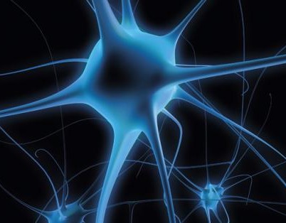
June 13, 2018 – Amyotrophic Lateral Sclerosis (ALS), better known as Lou Gehrig’s Disease, the condition that Stephen Hawking lived with for most of his adult life, causes paralysis as neurons in the brain lose their ability to communicate with the body. The disease kills 80% of those afflicted within two to five years after diagnosis. The disease is defined in three categories: sporadic, familial, and bulbar.
At the ALS Canada website, it states that more than 200,000 around the world are living with the disease. Of that total, 3,000 Canadians with 2 to 3 dying every day. The lifetime risk of getting ALS is 1 in 1,000.
Stem cell treatments for ALS are at the centre of most research seeking cures for the disease. One of these has led to the announcement of a new clinical trial focused on using human embryonic stem cells to propagate astrocytes, the most numerous cells found in the brain. Astrocytes are glial cells. The word glial is derived from the same root word for glue, and that’s exactly what astrocytes appear to be, the cells that hold together our brains and nervous system.
Astrocytes come in numerous subtypes called fibrous, protoplasmic, Layer-1, interlaminar, and varicose projection. Each serves a unique function. But all together astrocytes maintain our whole brain and neuronal metabolism providing mechanical and repair support while contributing to our neural plasticity, the ability of our brains to adapt in the face of changes. Astrocytes are also part of our human immune system acting as either pro or anti-inflammatory responders and thus are seen as key preventers of diseases of the brain.
So it would appear appropriate for researchers to look at astrocytes as a potential way to fight degenerative diseases like ALS. And that’s exactly what this clinical trial is all about.
Kadimastem is the biotechnology company that has been developing an embryonic stem cell therapy called AstroRx. In a recent study reported in the journal Stem Cell Research & Therapy researchers in Israel reported that transplanting healthy astrocytes derived from stem cells compensated for malfunctioning astrocytes in transgenic mice with ALS. The researchers manufactured large quantities of astrocyte progenitor cells from human embryonic stem cells. These were frozen and stored and then tested to see if they could produce healthy astrocytes. First, they proved it could be done in a laboratory. Then they moved on to animal studies injecting their AstroRx astrocyte cells into the spinal canals of their test mice. A regimen of two injections separated by 30 days proved to delay ALS onset while significantly improving motor performance and neurological test scores in treated mice. In a subsequent study, they administered larger dose load injections into test rats by lumbar puncture into the spinal canal. Results in the rat subjects were better than those in the test mice, delaying disease onset even further and slowing the decline in motor function. The study concluded that the method of treatment and the astrocytes derived from stem cells could be used in a human clinical trial.
That’s happening now at the Hadassah Ein-Kerem Medical Center in Israel. In the press release announcing the clinical results of the mice and rat studies, and the beginning of this new clinical trial, Michel Revel, Kadimastem’s chief scientist states, “In the research, we developed a method which enables the use of stem cells for the production of large quantities of healthy support cells, similar to those in the human brain, and the cells have been proven to possess several mechanisms of action for the protection of neuron survival.”
On the Canadian ALS website, there are a number of other clinical trials using various stem cell therapies to try and slow down or stop ALS symptoms from progressing. I’ll keep an eye on these and report back from time to time on their progress, and hopefully, their success.

But 1963 may not be the earliest date in which a cell exhibiting stem cell characteristics is mentioned. The term is mentioned in a 1932 medical journal, but the reference to what we know as stem cells today seems inappropriate. And then there are older references to stem cells back to 1905.
One thing is clear, however, we had little understanding of what these cells were and their potential.
Today we know about stem cells. The Mayo Clinic’s website calls them the “body’s raw materials,” cells from which all others can be generated. But what makes them so different from all those other cells in our bodies? Our bodies contain 200 known varieties of cells. Each, with the exception of blood cells, has a nucleus, mitochondria plus other organelles, a common chemical soup, and consistent structural features.
Yet there is no such thing as a typical cell. All cells pretty much do the same thing. They divide until their ability to do it stops. At that point, they die. We lose 96 million cells per minute but we also gain that same number in the same amount of time. The behaviour of each cell is determined by instructions given by genes located on chromosomes within the cell nucleus.
When we humans start out, our first cell is a fertilized egg combining DNA from both of our parents. The egg divides to eventually form a ball of cells we call an embryo. And the modern story of stem cells begins with what scientists first discovered about embryos. They contained cells that were non-specific but capable of being instructed to differentiate to turn into us, producing our bone, muscle, organs, brain, nerves skin, and nails. With all that differentiation leading to the 200 known varieties of cells we find in our bodies today, scientists overlooked one. Stem cells didn’t cease to exist when the embryo began to differentiate. They very much exist within all those differentiated organs and tissues that form us. You can find them in muscle, bone, organs, and fat. In fact, body fat may prove to be the best source of harvestable stem cells that we can use to cure so many of the chronic conditions and diseases we humans encounter.
Most of us when we think of stem cells, we think embryonic. That’s because that’s where the science of stem cells first started. To make embryonic stem cells researchers use a technique called somatic cell nuclear transfer. It involves manipulating donated eggs by removing the original DNA contained in the nucleus and replacing it with DNA from a donor adult cell. Cell division then follows until an embryo forms. The embryonic cells, about 100 of them on maturity, are then harvested and the embryo destroyed.
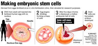
It is this use of human embryos that has created the controversy associated with stem cell research. For those who aspire to the belief that life begins at conception, harvesting and destroying an embryo is seen as murder. Lobbying by religious groups soon led to restrictions in the science in the United States with only a limited number of embryonic stem cell lines allowed.
But the promise of these cells and their ability to differentiate into any type of tissue has kept research laboratories busy in places other than the United States where more than 300 existing lines of human embryonic stem cells being cultivated in Canada, Sweden, France, Spain, and the United Kingdom. These lab-grown embryonic stem cells represent one branch of stem cell research. And because the cell populations are artificially cultured in laboratories there are some significant problems that can happen.
Consider evolution, the engine that drives speciation. From reptiles came dinosaurs, mammals, and birds. How did that happen? Mutation. And mutation worked best when there were large populations of individual species who through breeding, living and dying further differentiated and survived.
Now apply that same observed principle to laboratory populations of continuously dividing limited lines of stem cells. Just like in an isolated population of animals with a limited amount of DNA to share, mutations can produce undesirable results. In the case of laboratory-grown stem cells, a cell meant to cure could because of undesirable changes kill.
In addition, growing a large enough population of embryonic stem cells to cure people is an expensive proposition. The science, therefore, needs to find a better way to use the potential regenerative capacity of these cells.
Finding a new source of stem cells is critical. And lo and behold they are there, in our bodies, lurking among all those differentiated cells which came after conception.
In Part 2 we’ll continue the story, describing how readily available stem cells existing within us represent a medical revolution in tackling a wide variety of diseasee challenges from autoimmune to cardiac, hepatic, pancreatic, gastrointestinal, respiratory, neurodegenerative, ophthalmologic, spinal cord, skin, osteo, and more.
There have been many attempts over the years to create synthetic blood, a product that mimics blood’s essential purpose as the transporter of of oxygen and other nutrients throughout the body. But in every case no equivalent to natural blood has emerged.
Making blood from stem cells, however, would be a game changer. The goal would be to create the equivalent blood type, group O, rhesus negative. This is a type of blood that we define as the universal donor, a product suitable for all transfusion recipients. You see my blood can only be given to those who are also A Rh+ or AB Rh+. There are a lot of people on this planet with A Rh+ blood so in donating I can help them. There are very few with AB Rh+ and even so I can help them as well. But if all of this sounds a bit confusing here is quick blood type primer.
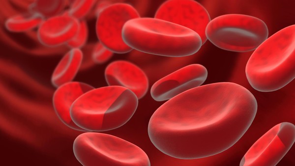
We have known since the early 20th century that not all blood is the same. That’s because the red blood cells each of us has contain different types of antigens. Antigens are sugars and proteins that can be found adhering to the surface of blood cells. Mismatched transfusions can trigger an immune system response. That’s why all who receive blood need to have their type identified before suitable donor blood can be dispensed. Our antigens are determined by our genetic makeup, our DNA. If our genes are not encoded for a particular sugar or protein then that antigen is absent from our blood cells. The absence or presence of these antigens determines the type of blood we have.
The identifying letters, “A,” “B,” and “O,” used here in North America and throughout much of the rest of the world (Russia and Eastern Europe use a numbering system) define the primary blood types. A secondary typing is known as “Rh.” And there are even more which we call subtypes, some of which are very rare. These are designated by the addition of a number beside the letter. So if you have your blood typed or if you choose to donate blood, you will be told that your blood is one of the following: A Rh+, A Rh-, B Rh+, B Rh-, AB Rh+, AB Rh-, O Rh+, O Rh-. Further testing may yield a sub-type such as A1, A1B, A2, A2B, and B1. These are among the 200 blood groups that modern science has identified.
Remember at the beginning of this posting I said that your blood type can tell you a bit about your origins. Here is a bit of blood geography lesson.
“A” blood type populations tend to be found in Central and Eastern European populations with one exception. The Blackfoot tribe in the United States happens to be predominantly type “A.”
“B” blood type populations are most commonly found in Asia, particularly China and India.
“AB” types are least common and found mostly in Japan, Korea and China.
“O” types, the most common are found in African, Hispanic and North American tribal populations.
The secondary blood typing “Rh,” which stands for rhesus factor, was first discovered in 1940 while testing inter-species blood transfusions between rhesus monkeys and rabbits., Rh+ is far more common than Rh-. Caucasian populations (European) tend to have the highest incidence of Rh- blood while Asian, Hispanic, Black, and North American aboriginal populations are predominantly Rh+.
Why is “O Rh-” considered the universal donor. Because both the “O” and the “Rh-” stand for the absence of the antigen proteins and sugars that cause immune responses.
Mismatching blood types has always had deadly consequences. Today such incidents are fairly rare. For example, in 2008 in the United States, 24 million transfusions were performed with 54 transfusion-related fatalities. In 2011 the number of transfusion deaths was 69. Before the turn of the 20th century mismatched blood deaths were common.
The goal of researchers in the United Kingdom, who have been working on producing blood from stem cells are trying to create a match to “O Rh-” for obvious reasons. This type of blood could be used by anyone. The researchers have chosen to work with embryonic and induced pluripotent stem cells (iPSCs). Starting with the embryonic type they can produce limited quantities of universal donor blood. But using iPSCs gives them access to much greater production volume. So the plan is to develop universal donor blood eventually from iPSCs. After four years the results are showing considerable promise with the first trials which they call “in-man,” scheduled for late 2016. “In-man” means exactly what it says. These trials will involve transfusing blood made from stem cells into human recipients.
One of the researchers in the program is Marc Turner, medical director of the Scottish National Blood Transfusion Service, describes the challenges and goal that lie ahead for the team. “Producing a cellular therapy which is of the scale, quality and safety required for human clinical trials is a very significant challenge, but if we can achieve success with this first in-man clinical study it will be an important step forward to enable populations all over the world to benefit from blood transfusions.”
In the meantime, I’ll still be donating blood every 56 days or so.

Stem Cells
These life-changing undifferentiated cells are generated and harvested from 3 sources.
A human embryo in its earliest stages is called a blastocyst. It consists of two types of cells, an outer wall that becomes placental tissue and an inner wall of cells that eventually differentiate into all the tissues of the body. It is these latter undifferentiated cells that are potential tools for fighting disease and healing damaged tissue. This is a new field of reparative medicine done at the cellular level.
Stem cells can be reprogrammed. They have unique regenerative powers. Studying their growth is helping scientists to understand what causes birth defects. Currently researchers study stem cells for their potential to deal with a range of human diseases and problems including: stroke and traumatic brain injury, Alzheimer’s, Parkinson’s, blindness, deafness, traumatic wounds, ALS (amyotrophic lateral sclerosis), muscular dystrophy, diabetes, Crohns, cirrhosis of the liver, spinal cord injury, rheumatoid and osteoarthritis, heart disease, heart lesion repair after a heart attack, and many cancers. Researchers are even using stem cells to design and improve pharmaceuticals to target specific diseases and medical conditions.
Embryonic Stem Cells
We have experimented with embryonic stem cells since 1981 when scientists derived stem cells from the embryos of mice. By 1998 we discovered how to do this with human embryos. Only embryos discarded from in vitro fertilization procedures were used but nonetheless controversy soon erupted from religious groups that saw using these embryos as the equivalent of murder.
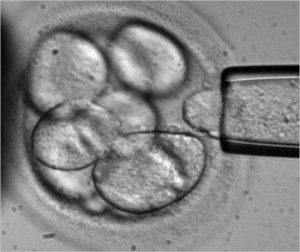
Embryonic stem cells can be cultured in a laboratory to divide while retaining their undifferentiated state. Called Pluripotent embryonic stem cells, they form stem cell lines which can be frozen and sent to laboratories and medical facilities for further research and testing.
In studying these cells researchers learn about human development and the complex processes involved when undifferentiated cells begin to differentiate to form various body tissues. This will help in understanding the mechanism that can lead to abnormal cell division and differentiation resulting in cancer and birth defects.
Somatic or Adult Stem Cells
Less controversial than embryonic, somatic (meaning cells not harvested from embryos) stem cells consist of undifferentiated cells found within body tissue that mostly contains differentiated cells. Somatic stem cells exist in organs and tissue in a caretaker role, providing maintenance and repair. We have been working with a form of somatic stem cells for some time. Probably the most well-known use is the procedure known as bone marrow transplantation used to treat leukemia, where hematopoietic stem cells are harvested from a donor and transplanted into a recipient.
Somatic stem cells have already been used to show their potential to create an endless supply of blood for transfusion allowing patients to bank cells that can be generated as needed eliminating the need for blood donors.
As researchers discover somatic stem cells in many more organs and body tissue their usefulness for other types of transplants may lead to cures for Parkinson’s, Alzheimer’s, heart disease, and many other intractable medical conditions.
Induced Pluripotent Stem Cells
Known in short form as iPSCs, these are adult tissue and organ cells that have been genetically reprogrammed to assume an undifferentiated embryonic stem-cell state. First “invented” and identified through the genetic manipulation of mouse cells in 2006, by 2007 human iPSCs had been harvested and cultured in laboratories.
Currently iPSCs are used for drug testing and modeling of diseases. Genetically altering an adult cell is no simple task. Using viruses as delivery agents, altered genetic materials get introduced into the cell. Unfortunately in some animal studies the viruses when introduced have caused cancer. We are not, therefore, ready to deploy iPSCs for therapeutic purposes in human studies. Researchers are exploring non-viral methods of delivering new instructions. Once they have mastered this the potential for therapies is enormous.
iPSCs can overcome tissue rejection. They can be used to repair and grow healthy tissue. This is the science of cell-based therapy and has the potential of replacing organ and tissue transplants. iPSCs can be created from a human source, cultured and directed to differentiate into specific cell types to treat diseases such as diabetes creating healthy insulin-generating cells in the pancreas, or repairing brains suffering from Alzheimer’s and Parkinson’s, or grow new heart muscle and blood vessels to cure heart disease, or repair immune systems that lead to osteo and rheumatoid arthritis. iPSCs have the potential to repair traumatic injuries such as spinal cord injury, or stroke, and regrow healthy skin to repair burns.
Stem Cell Therapy in the Medical Mainstream
How far are we away from seeing stem cell therapy in the medical mainstream? Today stem cells are being used to treat a small number of diseases and largely in clinical trial only. The most common use is blood stem cell transplantation for treating cancer. The PISCES study (Pilot Investigation of Stem Cells in Stroke), a fully regulated clinical trial of neural stem cell therapy for stroke patients started in 2010 in the U.K. In the United States clinical trials have started using embryonic stem cells differentiated into retinal cells to treat macular degeneration.
Soon these studies will be joined by many more. But first cell-based therapies need to move out of research laboratories. into many more clinical trials and then produce documented and consistent results to show that manipulated stem cells can be differentiated for transplantation consistently. That means creating enough cell volume before differentiation and controlling differentiation to create desired tissue types that match individual patients. That means ensuring that the tissues created from stem cells function the way they are supposed to for a lifetime and that the human host doesn’t experience rejection or develop any diseases resulting from transplanted cells.
We should be there if not within this decade then by 2030.
]]>


