Hai Zhang, a professor of restorative dentistry at the University, along with several colleagues has found a way to generate ameloblasts. What are they? Ameloblasts are one of two cells that exist in human embryos responsible for the formation of our teeth. The other cells are called odontoblasts—the former secrete enamel, the latter dentin.
The process of tooth development is called odontogenesis. The two cells mentioned above are critical to tooth formation. Enamel keeps our teeth surfaces hard and strong throughout our lifetimes. Mineralization of teeth begins early in embryonic development. Dentin precedes enamel production, both critical to giving us a healthy set of choppers.
But life has its travails. What we eat and chew causes the enamel surfaces of our teeth to wear thin, crack and decay. And because it is embryonic cells that form the enamel and then die out once our teeth have been produced, we have no biological means to repair them.
Researchers decided to take on the task of trying to create these lost before-birth enamel-producing ameloblasts. To do they used single-cell combinatorial indexing RNA sequencing or sci-RNA-seq. This technique reveals which of the genes in a cell are active in sequence during development.
The RNA molecule is the letter carrier of the cell while genes are the letter writers. Hence the term messenger RNA or mRNA (a term for which many of us have become more familiar from mRNA COVID vaccines), the molecule that carries the instructions to assemble proteins which serve as the letter reader and construction crew in our analogy. Changes om levels of mRNA differ during stages of embryonic cell development which determines which of our genes are sequentially activated.
What the University of Washington researchers were able to do is track each gene activation stage and create a roadmap to develop ameloblasts from undifferentiated stem cells using a software tool called Monocle. The software was able to predict the biochemistry sequence to test on stem cells. They exposed the cells using computer-designed proteins and chemicals. The latter activated different genes in sequence to create the final roadmap. While doing the work on ameloblasts they uncovered another cell type, subodontoblasts which may precede the development of odontoblasts.
The end result was the stem cells were induced to create multicellular mini-organs called organoids which resembled the structures seen in developing human teeth. Now the team is hoping to improve the process to make enamel as durable as that found in a healthy mouth and develop methods for applying the technique to fill cavities and do other enamel restoration. Imagine a future where we have stem cells activated to fill cavities, do other enamel repair, and even replace lost teeth.
The results of the current project were published last week in the science journal, Developmental Cell.
]]>

Today a dental laboratory represents a significant add-on cost to any dental practice. Every crown, bridge, implant or model a dentist uses is produced by them. The costs are factored into every dental bill patients receive and they are not inexpensive. when I had a crown done a few years ago, the total cost for all the work amounted to well over $2,000.
The dental laboratories employ skilled labor to produce crowns, dentures, partials and full sets of false teeth. The cost to an individual dental practice, depending on the volume of work, can amount to $100,000 in added overhead per year. And if a dentist takes the laboratory work in-house, the one-time cost of the equipment to do the work could easily reach $100,000 using traditional technologies.
But not anymore. The top-model 3D dental printers today sell for about $20,000 including a supply of materials. That’s one-fifth the cost of outsourcing the work to a dental laboratory. And when you consider that the cost of 3D printers is dropping to the point where they are becoming almost a commodity, the incentive for dentists to invest in them seems almost irresistible.
What can a dentist do with one of these 3D printing technologies in house? Here are just a few applications:
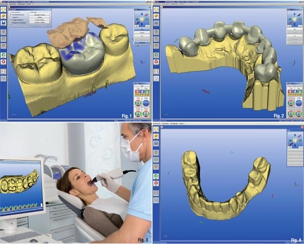
In a recent report on CNBC the dental sector is cited as the one most likely to “mass uptake” 3D printing technology growing 515% over the next decade. And ID TechEx in a recent forecast predicted that sales of 3D printers to the medical and dental sector will top $868 million US by 2025.
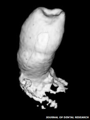
Researchers at the King’s College London, UK, in the Dental Institute have just published new findings in the current issue of Journal of Dental Research describing their experiments with mice to isolate adult human gum tissue cells and combine them with mesenchyme mouse cells, the cells in mammals that form teeth. The scientists once they had cultured a sufficient number of the cells then implanted them into a mouse jaw. The result, hybrid human-mouse bioengineered teeth containing dentine and enamel.
Professor Paul Sharpe, who led the team, stated, “‘Epithelial cells derived from adult human gum tissue are capable of responding to tooth inducing signals from embryonic tooth mesenchyme in an appropriate way to contribute to tooth crown and root formation and give rise to relevant differentiated cell types, following in vitro culture. These easily accessible epithelial cells are thus a realistic source for consideration in human biotooth formation. The next major challenge is to identify a way to culture adult human mesenchymal cells to be tooth-inducing, as at the moment we can only make embryonic mesenchymal cells do this.”
]]>

