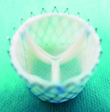
The effort is part of the European Union-funded LifeValve project, aimed at replacing mechanical and preserved animal tissue valves which are the current standard. The medical team cultures the heart valves from human cells. Design of the valves is done using computer simulations based on detailed imaging of a patient’s heart and outflow tracts to ensure that what is regenerated can be implanted without fitting problems.
Initially, the team used computer simulations to help understand how to culture human cells and grow them into a heart valve. They chose sheep for their animal models and were able over time to come up with accurate bio-engineered valves that could be implanted successfully. And now they have done it with the first successful customized implants in living sheep.
Why is this development so significant?
On a personal note, my daughter was born with a complex congenital heart defect called Tetralogy of Fallot with Absent Pulmonary Valve. She has had two open-heart operations and a third procedure where a valve was inserted into her pulmonary artery via a catheter. All three valves were cryogenically preserved dead animal valve tissue. My daughter faces a lifetime of these periodic valve replacements because dead tissue cannot regenerate itself.
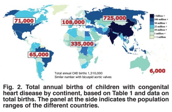
Worldwide the incidence of congenital (meaning born with) heart disease (CHD) is 8 to 12 for every one thousand live births, amounting to over 1.3 million globally each year. This worldwide phenomenon knows no boundaries occurring in populations everywhere with only minor variation by country in CHD type.
Structural repairs for most CHDs involves patching holes in the heart, altering arterial structures to improve blood flow either through the lungs or to the bodies extremities, and replacing missing or damaged valves. The human heart has four and depending on the type of CHD, any one or more of these valves could require a replacement implant.
With current practice using dead animal tissue valves, CHD patients undergo repeated surgical interventions throughout their lives. That’s because the valve flaps wear out, or calcify to become less flexible and restrict arterial blood flow. The alternative surgical prosthetic, mechanical valve implants, may last a lifetime, but require CHD patients to stay continuously on anticoagulants to prevent blood clots. But growing a heart valve with the CHD patient’s own human cells would eliminate the problems attendant to both current practices.
The biggest challenge is no longer in the growing but in the custom fitting of what is grown. There is no one size fits all solution. But it appears that the work being done in Zurich may soon be reflected in the first human clinical trials giving children born with congenital heart disease a much better life requiring fewer surgical interventions. Eventually, the bio-engineering of heart tissue using a patient’s own cells could heal the hearts of all children with CHD.
The results from Zurich have been published in the journal, Science Translational Medicine, May 9, 2018 issue, in an article entitled, “Computational modeling guides tissue-engineered heart valve design for long-term in vivo performance in a translational sheep model.”
We were lucky to some degree. Our daughter survived multiple bouts of pneumonia, two open-heart surgeries, and a pulmonary valve implant done by cardiac catheterization. Today she is 33 and married. Her prognosis is good but that doesn’t mean she won’t need further surgical interventions. And it doesn’t mean that she is not susceptible to respiratory infections and diseases that can compromise both heart and lung function.
She is an artifact of a time and place when the science and technology to combat congenital heart disease was still about cut and cover. But this is changing.
Enter Gene Editing Tools
The future for children like my daughter may soon be far different because of a tool called CRISPR/Cas9, a gene editing technology that I have talked about in the past at this blog site. CRISPR is the most well known of a number of gene editors capable of altering our DNA, as well as that of every animal and plant on the planet. It is helping to create disease and pest resistant crops. It is eliminating disease carrying mosquitoes. And now in humans, it holds the promise of removing unhealthy genetic information and replacing it with healthy DNA sequences. The ultimate capability of this technology is the elimination of all congenital diseases by snipping out the bad and replacing it with the good.
In the latest headlines reported in the last couple of weeks we have learned of an experiment conducted in the United States that targeted malfunctioning genetic information responsible for a congenital heart condition known as hypertrophic cardiomyopathy. This disease is very different from what my daughter had. But it has something in common. The disease is related to gene mutations that cause cells in the body to become disrupted leading to abnormal growth. My father-in-law succumbed to this disease in his 97th year. He was one of the rare ones who didn’t show signs of the condition until he was in his mid-80s. At that time he was outfitted with a cardiac pacemaker to help alleviate the worst symptoms of the condition which causes the wall between the two lower chambers of the heart to thicken to a point where it no longer is supple enough to contract. Hypertrophic cardiomyopathy normally begins to show symptoms in the population of adolescents and young adults. When it does it tends to lead to sudden cardiac death.
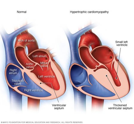
Like my daughter’s congenital heart condition, hyptertrophic cardiomyopathy (seen in the image above on the right) is associated with a range of gene mutations on one or more chromosomes. The research to date indicates 8 different gene mutations are linked to the disease. One of those genes is the MYBPC3 mutation which can be passed on to the child by either parent. It is defined as dominant inheritable trait which means only one copy is needed from a parent for the child to exhibit the mutated gene.
What does MYBPC3 do?
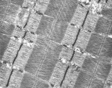
It provides important instructions that affect the contractions of the heart muscle. Specifically, it alters sarcomeres, the cells which make up the thin and thick filaments within the heart muscle (see image below). When the mutation is not present the sarcomeres work conjointly to create normal contractions. But when the MYBPC3 mutation is present the protein carrying instructions responsible for creating the heart produce errors in the natural configuration of these cells. And although the mutation may be asymptomatic, in 30% of cases the heart muscle will thicken to a point where symptoms occur.
The latest research from Oregon Health and Science University, Portland, that was published recently in the journal, Nature, attempted an experiment where mutated MYBPC3 genes were targeted using CRISPR/Cas9 to substitute good genetic information for bad. A total of 58 human embryos not destined for implantation were targeted. The team successfully corrected the genetic information responsible for the defect in 42 of the embryos while not causing any unwanted genetic changes in the remainder of the genome. Normal copies of the MYBPC3 gene were implanted using the Cas9 protein which serves as the knife tool within CRISPR. First Cas9, working with the embryo’s own RNA guide molecules selected the place for editing the gene. Once the defective gene was excised, it was replaced with healthy genetic information. Upon completion, the cell’s machinery carried on as if nothing had happened.
Such a technology, if replicated in treating other congenital diseases at the embryonic stage, would be revolutionary. Then parents who were known carriers of harmful genetic mutations based on DNA analysis and a comprehensive family history could have doctors intervene at the embryonic stage to correct life-harming and life-ending defects.
Of course, the dark side of speculation by journalists covering the story led to headlines that declared “designer babies” are in our future. Parents with perfectly normal medical histories and DNA exhibiting little in the way of harmful mutations could decide they wanted a child with blue eyes even though both of them had brown. Or some could request doctors to alter other genetic information that could bestow on an unborn child improved physical strength or mental acuity.
But we are learning far more about the genetic mutations that lead to congenital heart disease and a recent study from Yale University sheds some light on this subject. As reported in the November 13 online edition of Nature, a deletion in the gene Galnt11 is associated with a birth defect that affects the left-right orientation of organs in a developing embryo. It does this because it affects the motility of the cilia, hair-like structures in the body seen in the image below that drive fluid around within the embryo. With the deletion the balance of motile and immotile cilia is altered and this causes improper embryonic development.
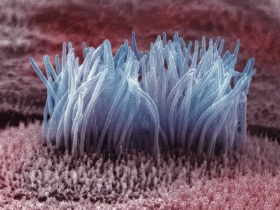
Improper orientation of developing organs is the end result. This can lead to a number of CHD conditions including holes developing in the septum of the heart, the wall that separates the left and right pumping chambers. Or reversal of the heart with the weaker pumping chamber pushing blood to the aorta rather than the pulmonary artery.
CHD today is the most common major birth defect with an average of 1 in 100 live births having some form of heart lesion. When first forming the heart begins as a symmetric tube that eventually loops around to form its four chambered array. The role of the cilia in ensuring proper left-right asymmetry has been known for some time. But now because of the Yale study we know of a particular gene mutation that interferes with the process.
Galnt11 joins a group of identified known genetic causes responsible for CHD. And what we are seeing is a preponderance of associated birth defects in 25 to 40% of children who receive a CHD diagnosis. For example 50% of children born with the genetic defect known as Trisomy 21 have a range of heart defects affecting the pumping chambers and septum separating them. Trisomy 13, another genetic defect, is associated with septal defects, as well, in 80% of CHD cases. About one-third of females born with a missing chromosome, known as Monosomy X, suffer from left-side malformations of the heart leading to diseases such as aortic stenosis or hypoplastic left-heart syndrome. About half of males born with Klinefelter syndrome, a condition where the child is born with 47 instead of the normal 46 chromosomes, suffer from patent ductus arteriosus, a condition where the pre-birth circulatory ducts do not close in the normal fashion leading to pulmonary hypertension, and ASD, a condition where holes appear in the septum that separates the upper two heart chambers. The latter condition if untreated eventually increases blood pressure in the lungs leading to hypertension as well.
Where are discoveries like Galnt11 taking us? In finding genetic origins to diseases we are beginning to unlock the mechanism of DNA. This is giving us insight into new methods of treatment and intervention. The emerging field of pharmacogenetics is leading to the development of targeted treatments that can insert corrected genetic information using synthetic proteins or adenoviruses containing missing genetic information. We are only now in the last two decades starting down this amazing path which will lead to early intervention, even in utero, so that babies will no longer be born with heart and other birth defects.
Insects in Your Diet an Almost Certain Future
Today 2 billion humans supplement their daily diet with insects. Didn’t know that did you? The technical term for eating these creatures is entomophagy and a United Nations study outlines the benefits derived from this protein source as one way for humans to deal with forecast growth in our numbers over the next three decades. Some highlights from the report:
So bon appetit!

This Story Gives A Whole New Meaning to the Words “Power Plants”
Researchers at the University of Georgia in Athens have published a paper in the journal Energy & Environmental Science, describing a method to interrupt photosynthesis to capture electrons before the plant uses them to create sugars. The technology separates thylakoids, a structure within plants responsible for capturing and storing energy from sunlight. They do this by manipulating the proteins within the thylakoids using carbon nanotubes which act like electrical conductors. This produces an electrical current. The field is bio-photovoltaics and we may see it used initially to power small remote sensors. But as the it improves we may see plant photosynthesis powering our appliances (see image below), our homes, cars and buildings.

Offer of $250,000 Prize for Human Powered Helicopter Flight Remains Unclaimed
The Igor I. Sikorsky Human Powered Helicopter Competition has been around since 1980. Set up by the American Helicopter Society the initial prize offered amounted to $20,000. Now 33 years later the pledge has grown to a quarter million U.S.
To win the money you have to build a human-powered helicopter that can takeoff from level ground, hover for one minute within a confined amount of space to show stability, and achieve 3 meters (approximately 10 feet) in altitude at some point during the flight.
The device has to be heavier-than-air so no helium or hydrogen balloons allowed. It must have a rotary wing configuration capable of vertical takeoff. It has to have at least one person on board for the entire flight including during initial rotor acceleration to obtain lift.
The device can have no additional assistance from technology that stores energy (no batteries) to enhance performance. Of course that doesn’t include the rotor used for lift or rotating devices that provide navigation and stability control. Nothing can be jettisoned from the craft during flight.
Last summer new rules were added to allow for a flywheel addition to the drive system as long as it is not used to transfer energy to the rotors.
Right now there are three teams chasing the prize. These include a team from University of Maryland, with Gamera II, the Upturn II from Cal Poly San Luis Obispo, and the Atlas (seen in the image below) from AeroVelo at University of Toronto.

New Study Reveals Non-Inherited Gene Mutations Responsible for Many Heart Defects
I have a particular and personal interest in this story because congenital heart disease (CHD) runs in my wife’s family history and after our daughter was born with a complex heart defect, we underwent genetic counseling that indicated we had a 6% chance of recurrence in a future child. So when I read about this latest study that states at least 10% of severe CHD occurrences come from mutations absent in parents, I was intrigued.
The research comes from Yale School of Medicine and was published on May 12 in Nature and implicates a common pathway involving several hundred genes that contribute to children being born with severe heart disease. The study involved studying 1,800 individuals and several hundred mutations. The study describes how proteins orchestrate the activation of genes in the developing fetus. Mutations in these genes then set off the incidents of CHD. The study also found some interesting overlaps with genes that caused CHD and autism. The researchers believe that what they have discovered is common pathways for many congenital diseases.

NASA Continues to Hype a Plan for a Human Mission to Mars – What Could Go Wrong?
In an online poll about going to Mars, conducted by The Engineer, a UK site focused on engineering job opportunities and industry news, results showed that more than 60% of respondents felt that landing would turn out to be the toughest of all the tasks. Other choices included building shelters to protect from radiation (22%), finding water (11%), preventing ion rocket failure over the duration of the flight (5%), and developing lightweight, folding photovoltaics to provide energy (1%).
The NASA plan for Mars recognizes that all of the concerns described in the poll represent hurdles to overcome. But by far the biggest is getting a human occupied spacecraft from Earth to Mars through Deep Space while ensuring the voyagers within are sufficiently protected from both cosmic rays and any other dangers they may encounter along the way. Current technology that can successfully put a robot like Curiosity on the planet doesn’t meet these challenges. The propulsion systems in current use mean a human voyage to Mars would take up to six months. Closed environment technology for sustaining human life for that period of time has yet to be developed and tested here on Earth. And so far any landings on Mars have always been a one-way trip. Although Mars One may intend one way trips for its human voyagers that is not in the cards for NASA.

A Postscript
So that’s the headlines I chose to share this week. I hope you found them interesting and as always look for your questions and comments. I should be back talking about our technological and scientific progress in the 21st century before next Friday, so expect to see some fresh headline stories then.
As always, thanks for dropping by.
– Len Rosen
For those of you who know me, CHD is a subject near and dear to me because of my daughter. She was born with a rare heart lesion and has had multiple surgical corrections throughout her life. That’s one of the reasons I have been studying and writing about biomedicine for so many years.
The new procedure comes from one of the hotbed facilities doing CHD research, Boston Children’s Hospital in Boston Massachusetts. The surgeons and cardiologists there have been working to find a way to let the body help itself in repairing the rare condition known as Hypoplastic Left Heart Syndrome (HLHS) seen in the image below.
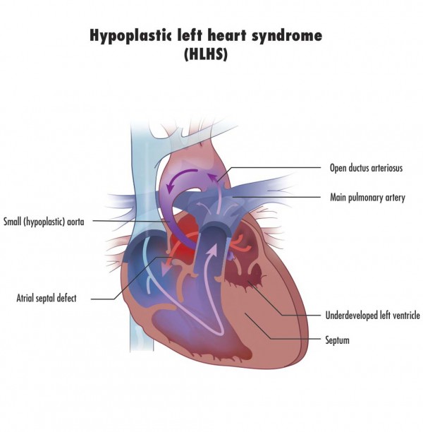
Children born with this defect suffer from an underdeveloped, almost non-existent left-ventricle. Normally that venticle serves as the major pumping chamber of the heart pushing blood through the aorta to the rest of the body. But in HLHS children the chamber is malformed. When the child is in utero the condition is not life threatening. But once born HLHS without intervention is a death sentence. HLHS is very rare with maybe 1,000 children born in the United States and 100 in Canada each year. Early surgical intervention is the only chance these children have for surviving days let alone weeks, months or years.
When a HLHS baby is born they are immediately compromised. Early intervention attempts to keep the holes that exist in utero between the major blood vessels and the two upper chambers of the heart open. Normally these close within a few hours after birth. With the holes open there is at least some circulation of blood to the body’s major organs.
Surgical intervention prior to this new protocol has involved creating temporary fixes. The first is called the Norwood Procedure which turns the right ventricle used to pump blood to the lungs into a single pumping chamber pumping blood to both the lungs and the rest of the body. Using the right ventricle this way has drawbacks. The right side of the heart is less muscular than the left because the circulatory demands on it are far less than the left. In turning it into a dual-purpose pumping chamber the muscle gets highly stressed and eventually fails. That’s because there isn’t enough muscle to do both jobs.
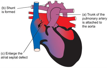
If a child survives for a period of time after a Norwood then other procedures can be done to try and create as close to a permanent fix as possible. But ultimately the preferred treatment for any HLHS child is to find a donor heart. Transplantation comes with its own challenges including donor availability and rejection management and therefore is considered a last resort. Surgeons would far prefer to use a body’s original parts whenever possible to affect a cure. That’s why finding a way for the body to self heal is considered ideal.
So the team at Boston Children’s set out to do just that, find a way to stimulate the left ventricle to grow from its hypoplastic state to a near normal size. Their newly developed surgical protocol is a technique that involves directing blood not away from but to the underdeveloped left ventricle and by doing this triggering the ventricle to grow so that it can eventually assume its normal function.The doctors who have pioneered this technique recently reported preliminary results in the Journal of the American College of Cardiology. They selected 34 patients for the new procedure and reported that 12 of them now have two working ventricles.
What’s involved with the protocol? Multiple surgical interventions. The first procedure alters the circulation of blood in and around the heart to stimulate left ventricle growth. Secondary procedures follow to repair or replace malformed or occluded heart valves, close off any holes between the pumping chambers, and reduce the amount of inelastic tissue in and surrounding the heart so that it can pump more efficiently.
Considering past results, even with multiple surgical procedures, this new protocol should give HLHS children a fighting chance at a full life.
]]>A Quick Primer on Heart Disease
Humans get two types of heart disease: congenital (that is born with) and acquired (disease that develops over time). Congenital tends to be structural. It can express itself in abnormal anatomy or physiology. Acquired begins with normal anatomy but physiology changes as the heart becomes diseased. Both can lead to heart failure which you can live with for a very long time or heart attacks which can lead to death.
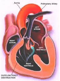
Structural defects to the heart and blood vessels in congenital heart disease can be repaired surgically, missing valves implanted and blood flow restructured using human grafts and artificial materials. Drugs can help regulate heart rhythm or improve the pumping efficiency. Pacemakers can help overcome structural problems that impact the electrical system that regulates heart rhythm.
Acquired, which represents the bulk of what we normally think of as heart disease, can also benefit from surgical intervention, drugs, pacemakers and other devices.
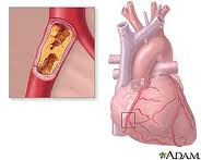
What causes heart disease? Lifestyle and inherited genes are the principal cause with 30 to 60% of the risk associated with the latter when it comes to acquired, rather than the former. Recent international studies identified 17 new genetic variants linked to increased heart disease risk. Five of these were associated with genetic regions that govern lipid metabolism processes, genetic controllers that impact the build up of fatty deposits in arteries. Ten others were in genes involved with other diseases and traits such as autoimmune diseases like Celiac and Type 1 Diabetes, cerebral and abdominal aneurysms, and lung cancer. What this means is there is no single genetic “smoking gun” that defines whether a person is likely to get heart disease in his or her lifetime. Instead we have a complex of genetic markers.
New Therapies for Repairing Bum Hearts and Blood Vessels
Stem Cells
In past blogs we have talked about stem cell therapy. Because stem cells have the potential to develop into a range of tissue types we can use them to regenerate and replace diseased tissue.
Cellular Dynamics International (CDI), is an American company located in Madison, Wisconsin, that is experimenting with the reprogramming of adult somatic cells to make them revert to a pluripotent state so that they can grow into any cell type needed for doing repairs to the body. These are induced pluripotent stem cells or iPS. Since 2008 CDI has used iPS cells to manufacture cardiomyocytes (heart muscle cells), freezing and sending them to pharmaceutical companies doing research on new drugs to treat heart disease.
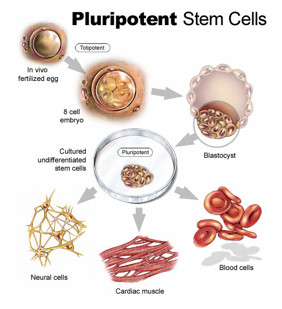
But iPS cells have a brighter future if we can work out some of the bugs. Those bugs include faint traces of chemical residue from the parent cells used to create the reverted cells. Those residues can lead to mutations and as we have already written in previous blogs, mutations can lead to runaway cancer cell proliferation.
You can create iPS cells from almost any human tissue source. If a patient with a damaged heart provides a tissue sample, the cells from the sample can be induced to revert to iPS cells. They then can be transplanted back into the patient at the location requiring treatment. iPS cells from the donor are not rejected because they are from the donor who is also the recipient. The iPS cells in their reverted state can take on the characteristics of those cells that are in proximity to them and start to divide. iPS cells in the heart can generate heart muscle. iPS cells in the spine can generate nerve cells. CDI charges pharmaceutical companies $1,500 for 1.5 million iPS cells. That’s not cheap but the potential for iPS as an alternative therapy to transplants makes this a desirable area of ongoing research.
In research being conducted in Bristol, England and Glasgow, Scotland, Doctor Costanza Emanueli, along with a team of scientists are studying a variety of approaches to treating heart attack victims including using embryonic stem cells to create blood vessel cells to inject into patients. The method of delivery is a skin patch.
Gene Therapy
Researchers are looking at ways to tweak DNA to repair injured hearts. This involves manipulating a variety of genes that impact muscle and blood vessel growth.
Dr. Emanueli’s team, mentioned in the previous section, is studying boosting levels of nerve growth factor (NGF) to improve heart attack survival rates. In studies with mice the death rate from heart attacks was reduced by half using NGF. The gene responsible for NGF was attached to a specially engineered virus and delivered into the hearts of mice. Mice that received the NGF gene showed much improved circulation and heart function. In previous studies NGF was known to regrow nerves but in this case it encouraged new blood vessels to grow inside the injured heart muscle.
In another study conducted at Harvard’s Beth Israel Deaconess Medical Center, the gene C/EPB-beta spurred the growth of cardiomyocytes. This study showed that a genetic trigger responding to physical exercise turned on a molecular pathway to get cells to start dividing. For a very long time cardiologists could not find evidence of cell division in adult heart cells. Hearts under stress or subject to exercise would swell but cell proliferation in these enlarged hearts was poorly understood.
Heart muscle enlargement, called hypertrophy, can be associated with disease or with what is referred to as “athlete’s heart.” In the study cardiomyocyte growth was attributed to molecular events rather than factors such as high blood pressure.
Reprogramming Heart Cells After a Heart Attack
Heart attacks in the past have caused irreparable damage to heart muscles. What causes them? The usual suspects are accumulations of deposits on the walls of arteries that supply the heart with blood. When an artery is partly to fully blocked oxygen and nutrients are cut off to the area next to the blockage. This kills the muscle near the blockage affecting the heart’s ability to beat. But what if the section of damaged heart muscle can regrow?
Professor Paul Riley, of Oxford University’s Department of Physiology, Anatomy and Genetics, and his team of researchers are looking at a layer of cells known as the epicardium. The epicardium is the inner wall of the sac of tissue known as the pericardium that surrounds the heart. The embryonic epicardium contains many types of cells used by the heart and circulatory system including blood vessels and heart muscle. But after birth the epicardium gets quiet and stops generating these tissues. Dr. Riley and his team looked at ways to switch the epicardium cell generation back on. In mice studies they achieved just that by administering doses of Thymosin Beta 4, a natural occurring protein, to stimulate the embryonic genes in the epicardium to create cardiomyocytes. The researchers found that administering Thymosin Beta 4 before a heart attack proved to stimulate faster regrowth of heart muscle tissue.
The team continues to hunt for other naturally occurring molecules that can stimulate heart muscle and blood vessel growth. Along with drug and interventional therapies to clear arterial blockages, administering these proteins pre-emptively could create a bank of healthy new heart muscle to speed recovery should an attack occur.

