In the world of internal medical robots HeartLander is a giant. Researchers have much smaller in mind when they conjure up micro-robot designs. These researchers develop robots on a nanoscale. We have broached this subject before in a blog and stated at the time of writing that there were no existing biomedical nanobots. That remains true but researchers at ETH Zürich have been building what they call micro-robots that are bacterial-scaled measuring lengths of between 5 and 15 nanometers. Called Artificial Bacterial Flagella or ABF for short, these devices swim like bacteria with corkscrew tails.
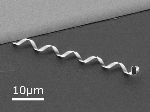
Made by depositing vaporized indium, gallium, arsenic and chromium onto a surface substrate a few atoms thick, the ABFs are patterned using lithography and etching. When thin sliced they naturally form the curled ribbon corkscrew you see in the picture above. The ABF head seen on the right contains a tri-layer film of chromium, nickel and gold. Nickel’s magnetic properties make it possible to use an external magnetic field to provide locomotion. The ABF uses helical propulsion (the same method of locomotion used by many bacterium) to swim through liquid at speeds of up to 20 nanometers at present with plans to increase it to 100 nanometers. Compare that to E. coli which swims at 30 nanometers per second.
To give ABFs autonomous power researchers are looking at thin-film rechargeable batteries. Currently these batteries can be manufactured and shaped with thicknesses of less than 50 nanometers. Chemical fuel sources may prove to be even more promising. An ABF running on energy it harvests from the blood would be capable of running indefinitely within a body harvesting glucose and oxygen to provide motive and computing power. Some researchers are looking at using bacteria as an ABF, modifying a living cell by attaching nanoparticles to it so that it can be mobilized for biomedical purposes.
What can ABFs do that current biomedical technology cannot?
Currently ABFs are confined to the research laboratory. But soon we will microscopically see these devices applied to all kinds of medical procedures reducing the need for surgical procedures, minimizing the side affects of cancer chemotherapy, and giving biomedical professionals a new set of tools for tackling conditions for which current medical practice has few solutions.
For those developing this technology it’s a question of tinkering with the power source to come up with the best way to deploy micro-robots.
]]>Assessment of the Physical Using Virtual Reality
What is assessment using virtual reality? It is the modelling of human movement in athletic and work environments to determine ways to mitigate risk to the body of injury both short and long-term. In the image below OpenSim software accurately models human movement to help understand the interaction of our muscles and skeletal structure, improve diagnosis related to injury, and provide early intervention to eliminate a potential health problem.

OpenSim was first made available as an open source platform to build, exchange and analyze musculoskeletal models and movement in the summer of 2007. Today it is widely used throughout the world by biomechanical engineers, computer scientists, robotics designers, neuroscientists, medical professionals, physical therapists, and sports physiologists. Fields of rehabilitation research include: stroke, spinal cord injury, cerebral palsy, prosthetics, orthotics, osteoarthritis and robotics-assisted therapy.
For children with cerebral palsy suffering from crouch gate, OpenSim allows doctors to assess whether a child is a suitable candidate for ham string muscle lengthening to correct this symptom that eventually can lead to permanent dependence on a wheelchair.
Using Virtual Reality to Treat Brain Trauma and Neuropsychological Problems
Virtual environment immersion is one of a number of treatment methods being deployed by medical professionals to deal with conditions like Post Traumatic Stress Disorder (PTSD). The United States military has had to deal with PTSD, concussion, and exposure to neurotoxins in its most recent wars in Iraq and Afghanistan. In the past tests for detecting brain trauma usually involved physical examination or written responses to a questionnaire. But researchers into this field recognize that cognitive testing using computers allows care givers to identify the nuances of different types of trauma and come up with specific therapies to help patients.

For veterans the video games provide multi-sensory environments similar to their wartime engagements. They do patrols on foot or in vehicles, are exposed to local native populations, some who are hostile, detonations from improvised explosive devices, shelling, bombing and grenades, and virtual gunfire. The games track neuropsychological measurements including eye movement, psychological and physiological changes, and systemic functions including cardiovascular and brain wave activity. For PTSD virtual reality provides exposure therapy, outperforming traditional exposure therapies dealing with PTSD symptoms.
Virtual reality therapy places a patient in immersive environments where phobias can be experienced. Patients become desensitized through repeated exposure in a safe, controlled atmosphere where psychologists measure physiological responses accurately and can treat any associated panic and anxiety. Virtual reality is being used to treat fear of flying, of driving, agoraphobia, social phobias and panic disorders, the trauma associated with accidents and PTSD.
The Center for BrainHealth at the University of Texas treats patients with Asperger Syndrome and Autism using virtual reality. The virtual world contains normal, everyday life experiences and settings. Patients practice social skills as they interact in virtual restaurants, stores, work settings and home. Practicing in these settings diminishes anxiety and fear and propagates greater independence. The technology allows the physician to increase complexity in the experiences to simulate real encounters and records the physiological responses to track progress. This rewires pathways within the brain of patients allowing them to lead more normal lives.
What is the Next Step in the Evolution of These Technologies?
As effective as simulations and video games can be, the development of holographic imaging will take us into a far more immersive environment in which even those we currently cannot restore to fully active normal life will be able to experience virtual space as if it were real. The question for all of us is “will we be able to tell the difference?” In future blogs we will return to this technology’s use in biomedicine as we explore its evolution through the mid-century and beyond.
]]>