Here goes……
Agriculture
This story bridges two subject areas, both agriculture and robotics. A robotics team at Kansas State University has built a prototype robotic tractor that when scaled up can be used by farmers for managing planting. A limited release of the first robotic planting systems is scheduled to go on sale this fall.

Biomedicine
This story appeared in April in Nature. Nanomaterials are playing a larger role in producing biomedical breakthroughs particularly in targeting treatment of conditions that have not responded well to traditional drug therapies or invasive procedures. In this case nanotechnology is making it possible to deliver drugs across the blood-brain barrier to treat cerebral palsy with the hope that animal studies will soon progress to human clinical trials.
Energy
Biofuels are increasingly appearing at local gas stations, in the wing tanks of commercial aircraft, and in diesel-powered ships. Bio refineries are springing up in both the Developed and Developing World. This article appeared in Aol Energy on May 22 and is an interesting read about biofuels, the food versus fuel debate and the technological advances being made.

The Environment
Kudos to the City of Los Angeles, the largest city in the United States to ban the use of plastic bags in grocery stores. If you need a bag you’ll be able to get paper at 10 cents per bag.
Outer Space
It would be hard to ignore the accomplishments this week of SpaceX with their successful launch of the Dragon spacecraft and its docking today with the International Space Station. We’ll be paying a lot more attention to commercial enterprises in space in future blogs.

Robotics/Artificial Intelligence
Creating robots that are anthropomimetic, that is they mimic humans in appearance, function and movement, is the goal of The Robot Studio. Read this article and look at the YouTube video that was posted in April about ECCEROBOT.

It is this capability that represents the significant breakthrough in this research. Scientists have been experimenting with synthetic DNA molecules for more than a decade. But in the research led by Philipp Holliger and Vitor Pinheiro, at the Medical Research Council Laboratory of Molecular Biology in Cambridge, United Kingdom, the research team was able to create synthetic DNA capable of duplicating allowing it to evolve. To achieve this the research team created six special polymerases that synthesized XNA from DNA and then copied the XNA back into DNA.

This is the essence of how evolution works. As DNA and RNA molecules replicate small errors over time appear. These errors alter the genetic code that underlies life here on Earth.
With XNA able to evolve through replication researchers can begin to understand how life started. The fact that XNA can do what DNA and RNA do suggests that there may have been other life creating molecular systems that predated or competed with them. Imagine life starting either here on Earth or somewhere in the Cosmos with competing forms of self-replicating molecular structures duking it out for supremacy. Here on Earth DNA and RNA were the winners. Could there be an XNA elsewhere leading to entirely different life forms? The research suggests the evidence is there.
Short of potentially creating life from exotic biology is there a practical value to the research? By developing XNA medical researchers can begin to explore ways to use it in therapeutic medicine, particularly in the development of nucleic acid treatments with modified DNA sequences that are self sustaining to target diseases. Current research in this area has been unable to create molecular-based treatments because the natural occurring DNA and RNA molecules breakdown before reaching therapeutic targets. The work on XNA should allow biomedical researchers to develop new anti-angiogenic medicines. Currently there is only one and it is used to treat macular degeneration.
]]>
A Quick Primer on Heart Disease
Humans get two types of heart disease: congenital (that is born with) and acquired (disease that develops over time). Congenital tends to be structural. It can express itself in abnormal anatomy or physiology. Acquired begins with normal anatomy but physiology changes as the heart becomes diseased. Both can lead to heart failure which you can live with for a very long time or heart attacks which can lead to death.
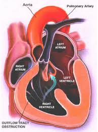
Structural defects to the heart and blood vessels in congenital heart disease can be repaired surgically, missing valves implanted and blood flow restructured using human grafts and artificial materials. Drugs can help regulate heart rhythm or improve the pumping efficiency. Pacemakers can help overcome structural problems that impact the electrical system that regulates heart rhythm.
Acquired, which represents the bulk of what we normally think of as heart disease, can also benefit from surgical intervention, drugs, pacemakers and other devices.
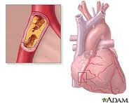
What causes heart disease? Lifestyle and inherited genes are the principal cause with 30 to 60% of the risk associated with the latter when it comes to acquired, rather than the former. Recent international studies identified 17 new genetic variants linked to increased heart disease risk. Five of these were associated with genetic regions that govern lipid metabolism processes, genetic controllers that impact the build up of fatty deposits in arteries. Ten others were in genes involved with other diseases and traits such as autoimmune diseases like Celiac and Type 1 Diabetes, cerebral and abdominal aneurysms, and lung cancer. What this means is there is no single genetic “smoking gun” that defines whether a person is likely to get heart disease in his or her lifetime. Instead we have a complex of genetic markers.
New Therapies for Repairing Bum Hearts and Blood Vessels
Stem Cells
In past blogs we have talked about stem cell therapy. Because stem cells have the potential to develop into a range of tissue types we can use them to regenerate and replace diseased tissue.
Cellular Dynamics International (CDI), is an American company located in Madison, Wisconsin, that is experimenting with the reprogramming of adult somatic cells to make them revert to a pluripotent state so that they can grow into any cell type needed for doing repairs to the body. These are induced pluripotent stem cells or iPS. Since 2008 CDI has used iPS cells to manufacture cardiomyocytes (heart muscle cells), freezing and sending them to pharmaceutical companies doing research on new drugs to treat heart disease.
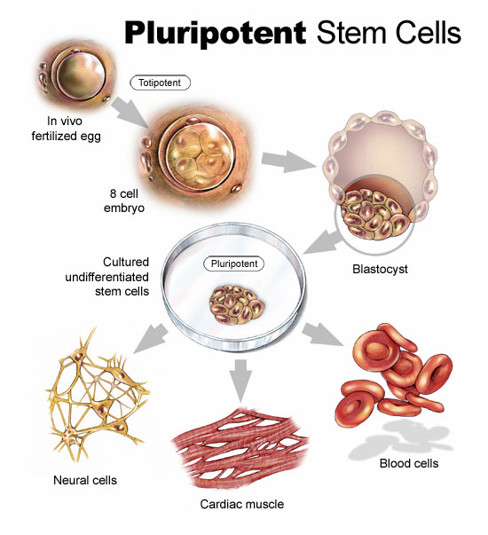
But iPS cells have a brighter future if we can work out some of the bugs. Those bugs include faint traces of chemical residue from the parent cells used to create the reverted cells. Those residues can lead to mutations and as we have already written in previous blogs, mutations can lead to runaway cancer cell proliferation.
You can create iPS cells from almost any human tissue source. If a patient with a damaged heart provides a tissue sample, the cells from the sample can be induced to revert to iPS cells. They then can be transplanted back into the patient at the location requiring treatment. iPS cells from the donor are not rejected because they are from the donor who is also the recipient. The iPS cells in their reverted state can take on the characteristics of those cells that are in proximity to them and start to divide. iPS cells in the heart can generate heart muscle. iPS cells in the spine can generate nerve cells. CDI charges pharmaceutical companies $1,500 for 1.5 million iPS cells. That’s not cheap but the potential for iPS as an alternative therapy to transplants makes this a desirable area of ongoing research.
In research being conducted in Bristol, England and Glasgow, Scotland, Doctor Costanza Emanueli, along with a team of scientists are studying a variety of approaches to treating heart attack victims including using embryonic stem cells to create blood vessel cells to inject into patients. The method of delivery is a skin patch.
Gene Therapy
Researchers are looking at ways to tweak DNA to repair injured hearts. This involves manipulating a variety of genes that impact muscle and blood vessel growth.
Dr. Emanueli’s team, mentioned in the previous section, is studying boosting levels of nerve growth factor (NGF) to improve heart attack survival rates. In studies with mice the death rate from heart attacks was reduced by half using NGF. The gene responsible for NGF was attached to a specially engineered virus and delivered into the hearts of mice. Mice that received the NGF gene showed much improved circulation and heart function. In previous studies NGF was known to regrow nerves but in this case it encouraged new blood vessels to grow inside the injured heart muscle.
In another study conducted at Harvard’s Beth Israel Deaconess Medical Center, the gene C/EPB-beta spurred the growth of cardiomyocytes. This study showed that a genetic trigger responding to physical exercise turned on a molecular pathway to get cells to start dividing. For a very long time cardiologists could not find evidence of cell division in adult heart cells. Hearts under stress or subject to exercise would swell but cell proliferation in these enlarged hearts was poorly understood.
Heart muscle enlargement, called hypertrophy, can be associated with disease or with what is referred to as “athlete’s heart.” In the study cardiomyocyte growth was attributed to molecular events rather than factors such as high blood pressure.
Reprogramming Heart Cells After a Heart Attack
Heart attacks in the past have caused irreparable damage to heart muscles. What causes them? The usual suspects are accumulations of deposits on the walls of arteries that supply the heart with blood. When an artery is partly to fully blocked oxygen and nutrients are cut off to the area next to the blockage. This kills the muscle near the blockage affecting the heart’s ability to beat. But what if the section of damaged heart muscle can regrow?
Professor Paul Riley, of Oxford University’s Department of Physiology, Anatomy and Genetics, and his team of researchers are looking at a layer of cells known as the epicardium. The epicardium is the inner wall of the sac of tissue known as the pericardium that surrounds the heart. The embryonic epicardium contains many types of cells used by the heart and circulatory system including blood vessels and heart muscle. But after birth the epicardium gets quiet and stops generating these tissues. Dr. Riley and his team looked at ways to switch the epicardium cell generation back on. In mice studies they achieved just that by administering doses of Thymosin Beta 4, a natural occurring protein, to stimulate the embryonic genes in the epicardium to create cardiomyocytes. The researchers found that administering Thymosin Beta 4 before a heart attack proved to stimulate faster regrowth of heart muscle tissue.
The team continues to hunt for other naturally occurring molecules that can stimulate heart muscle and blood vessel growth. Along with drug and interventional therapies to clear arterial blockages, administering these proteins pre-emptively could create a bank of healthy new heart muscle to speed recovery should an attack occur.
What is an Autoimmune Disease?
Autoimmune diseases are inflammatory response diseases caused by our immune system attacking us. They are non-discriminating and can be found in almost every organ and system in the human body. They tend to be chronic conditions continuing throughout the lifetime of the individual. There can be periods of remission followed by reappearance of symptoms. They tend to strike women more than men. Hormones may play a role. But there are some autoimmune disorders that are more prevalent in men such as Type 1 Diabetes.
How significant is the problem? It is estimated that 24 million Americans and 5 percent of the population in Western countries suffer from autoimmune diseases. Compare that to cancer at 12 million Americans and you begin to see the enormity of the problem.
There appears to be a correlation between our genetic inheritance and autoimmune diseases with children tending to develop diseases that appear in parents, and close relatives exhibiting common autoimmune diseases within extended family groups. Multiple sclerosis represents one of the most common autoimmune diseases that tends to run in families.
What confounds the medical field today about autoimmune diseases is finding the smoking gun or guns, the direct causal link that triggers the immune system response. Lots of circumstantial evidence points to viruses, chemical exposure, foods and environmental toxins but doctors and researchers continue to investigate what triggers these diseases. Having a family member who continues to experience an autoimmune disease has made studying this subject very personal to me.
Autoimmune diseases attack every part of the body. The American National Institute of Health identifies at least 80 different autoimmune diseases that include many familiar conditions you probably never thought were caused by our immune system. See how many you know from this partial list.
Today’s Autoimmune Controls Create Their Own Problems
Currently autoimmune disease treatment is all about reducing or reversing symptoms. In the case of diabetes we treat sufferers with insulin rather than restore pancreatic cell function. In the case of many of the inflammatory responses from autoimmune diseases we treat patients with a cocktail of drugs that include prednisone and other corticosteroids, anti-inflammatories like aspirin, acid blockers, antihistamines and immune suppression drugs (the kind used to stop rejection in organ transplants). In many cases the side affects of these drug cocktails proves to be worse than the diseases.
HLA, T and B, Autoimmune Diseases and a Potential Cure
When scientists and researchers talk about the immune system the letters in the above title become part of the conversation.
What is HLA?
What does T refer to?
What is the B all about?
HLA stands for Human Leukocyte Antigen, the genes that reside on the 6th chromosome in our cells and that govern immune response. The proteins associated with these genes are called antigens.
The T and B refer to different types of leukocytes, the white blood cells that reside in our circulatory systems and marshal our response to infections. The T and the B refer to T-cells, and B-cells. In addition there are sub-categories to leukocyte groups. One is the T-regulatory cell or T-reg and researchers study it because these cells play a role in regulating the level of immune response by other leukocytes.
If we could develop a technology to stop specific unhelpful autoimmune reactions while allowing our normal immune response to deal with unwanted bacterium and viruses then we would be making a big leap forward in our ability to deal with autoimmune diseases. That’s exactly what researchers at the University of Calgary in Alberta, Canada, have been working on using nanotechnology as the delivery mechanism for a vaccine that cures Type 1 Diabetes and has implications for other autoimmune disorders.
Dr. Pere Santamaria works at the Julia McFarlane Diabetes ResearcherCentre in the University’s Faculty of Medicine.

Working with mice with Type 1 Diabetes, Dr. Santamaria and his team looked at stopping the autoimmune response that damages the pancreas leading to the condition. They studied the behaviour of specific T-cells responsible for the disease and the T-reg cells whose role is to inhibit the former from attacking healthy host tissue. Their goal was to strengthen the T-reg cells to stop the destructive autoimmune response. Using synthetic iron oxide nanoparticles and a cocktail of antigens from insulin producing cells, the team created a vaccine that could be directed at the autoimmune attack, strengthening the T-reg cells. The treatment not only restored normal blood sugar and insulin levels in the mice, it also prevented the onset of the disease. At the same time the vaccine treatment did not compromise the rest of the immune system.
The implication of using nanovaccines such as the one Dr. Santamaria’s team developed to treat other chronic autoimmune diseases is enormous. For those who suffer from one or more of that long list of autoimmune diseases described above, this is technology with great promise.
]]>When the genes in normal (somatic) cells mutate cell behaviour may change over time leading to cancer. Mutations are a normal part of the life of a cell. That’s because when cells divide they replicate their DNA but imperfectly.
In a previous blog we talked about telomeres, the ends of the DNA strands in our chromosomes and how the telomeres shorten over time with each cell division eventually leading to cellular death. But it is not just telomeres that change during cell division. Other parts of the DNA can get scrambled or lost. We call these changes mutations. Since mutations happen all the time why do some become cancerous while others remain benign?
Medical researchers suspect that specific mutations in sections of our DNA that regulate the cell life cycle, when accumulated over time, cause cancer. Mutations that govern other cellular functions appear to have no malignant implications.
Here are some additional facts about cancer.
As researchers study cancer they are discovering new ways to treat it and reviewing some old ideas that showed promising results in the past. Let’s look at where we were with cancer, where we are today, and where we will be in the near future in the 21st century.
Seeking a Vaccine that Cures Cancer
Does the name William Coley ring a bell? For most of you, probably not. Born in 1862, Dr. Coley, an American surgeon who decided to devote himself to finding a cure for cancer, was the first to note that exposure to an infection could arouse a person’s immune system to shrink tumors.
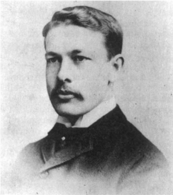
Dr. Coley studied sarcoma (bone cancer) at his New York hospital and identified a prior case of a German immigrant who had been operated on several times to excise a tumor in his left cheek. Each time the tumor regrew and after the final operation the remaining wound became infected. Surgeons were convinced that the man’s case was terminal but 4-1/2 months after he was discharged the tumor had vanished. When Dr. Coley studied the case he discovered that Streptococcus Pyogenes, a common bacterium that causes strep throat, was the source of the wound infection. The history of the case showed that the man had several outbreaks of fever and these outbreaks coincided with dramatic changes to the tumor. Dr. Coley concluded that the infection had saved the man’s life by stimulating his immune system to eradicate not only the bacterium but also the cancer.
Dr. Coley tested his theory on late-stage sarcoma patients first injecting them with live Streptococcus Pyogenes bacterium. The injections caused the tumors to shrink but in two cases the strep infections killed the patients. Dr. Coley then experimented with heat-treated bacterium injections. His first patient was a 16-year old boy suffering from a massive abdominal tumor. Known as Coley Fluid and later Coley’s Toxin, when injected into the tumor mass, produced the symptoms of an infectious disease (fever and chills) but not the full-blown illness itself. Repeat injections caused the tumor to shrink and eventually disappear. With no further cancer treatment the patient was discharged and survived another 26 years. Death was from a heart attack and not cancer.
Today the work of Dr. Coley continues at the Cancer Research Institute in New York. Founded by Dr. Coley’s daughter, the Institute studies how our immune system responds to cancer. In the 1970s doctors at the institute discovered that Bacille Calmette-Guerin or BCG could be used to treat early onset cancer of the bladder. The Institute has also studied cell proteins, called cytokines, and their immunotherapeutic effect on tumors.
Far from being Coley’s Toxin, seen by many in the medical establishment as quackery, current research is proving that Dr. Coley’s approach may lead to multiple cancer vaccines similar to the HPV cancer vaccine used to prime the immune system to kill human papillomavirus, a cause of cervical cancer.
Provenge is a vaccine developed by Dendreon, a company in Seattle. Designed to initially treat late-stage prostate cancer, Provenge is patient-specific. Cells from a patient are collected and then exposed to a chemical bath that contains cytokines that activate the immune system to attack the cancer. The cells are reinjected into the patient over the period of a month. Clinical trials on 512 advanced prostate cancer patients have been encouraging with 1/3 of the vaccinated patients remaining alive after 3 years. Plans are to introduce Provenge into earlier stage prostate cancer clinical trials.
We now know through the legacy of Dr. Coley that immunotherapy works. But what is the actual mechanism within our cells that leads to cancer? Research using baker’s yeast is yielding some exciting results.
Why Yeast Holds Clues to Curing Cancer
Saccharomyces cerevisiae is baker’s yeast, the yeast we humans have been using for milennia to make bread and fermented beverages. When a biologist, Leland Hartwell, decided to study cancer he chose yeast to help him model and understand the cell cycle.
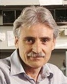
Hartwell was able to identify more than 100 genes directly involved in yeast cells that impacted life cycle. He called these Cell Division Cycle genes or CDCs. Hartwell identifed specific yeast genes responsible for different parts of the cell cycle and found similar characteristics in human cells. Since CDC genes either stimulate or inhibit cell division at very specific times in the cell lifecycle, Hartwell was able to identify the genes that didn’t operate in a normal manner. These included:
Any mutation to these genes could lead to what he described as driving with a stuck-accelerator and broken-brakes with no awareness that something has gone wrong resulting in out-of-control cell replication and the development of cancerous tumors.
What are the implications of this research in our search for cures for cancer in humans? Knowing that mutations in genes that control the lifecycle of our cells causes cancer should allow us to develop targeted therapeutic drugs specifically aimed at stopping runaway tumor growth. The tailor-making of drugs, called pharmacogenomics, should result in patient-specific cancer chemotherapy treatment without the side effects we normally associate with today’s treatments.
Having discovered the genetic mechanism that fuels cancer, the challenge is to find a way of delivering the cure and scientists may have discovered that answer. Read on.
RNA, Nanotechnology and Cures for Cancer
We’ve talked about Ribonucleic Acid or RNA in previous blogs. RNA interference or RNAi is a recent discovery and has enormous implications in delivering a cure for cancer. Why? Because RNAi can be used to silence the activity of specific genes within a cell. It does this by destroying messenger RNA or mRNA, the molecular messenger that carries coded information in genes to the protein factories needed to manage the cell’s lifecyle.
The challenge scientists faced was finding a way to dleiver RNAi to a target without it degrading. When RNA is normally injected into the bloodstream it quickly degrades. That’s where nanoparticles come in. American researchers have developed a vaccine that contains a nanoparticle drug that can deliver RNAi to cancer cells. Using a polymer that self-assembles to create the nanoparticle, and coated with a chemical that provides each particle with protection from binding to any cells it encounters within the bloodstream, the nanoparticles target surface receptors on cancer cells, penetrate and destroy them and cause minimal side effects to surrounding healthy cells.
It has taken 15 years to develop nanotechnology delivery systems. RNAi was discovered in 1998. What will we witness in the next 15 years? We are getting much closer to one of the Holy Grails of modern medicine, a cancer cure.
Our Biological Limits
In the latter part of the 18th century the average human lived 24 years. By the end of the 19th the average lifespan had doubled. In the second decade of the 21st century we are approaching a doubling again. Is there a biological limit?
We are the sum of our genetics. How long your parents lived may indicate how long you will live. But then again it may not. We know that altering genes can alter the lifespan of animals and plants we study in laboratories. We have doubled the lifespan of mice by splicing genetic material into their chromosomes.

In an earlier blog we described the structure of DNA, genes, base pairs and chromosomes. It may be helpful to click on the link provided as a quick refresher before reading further.
We have two DNA repositories in every cell in our body (blood cells not included). That DNA is found in the nucleus and mitochondria within the cell. The DNA organizes itself as chromosomes. When cells replicate the chromosomes divide and copy themselves. This is called mitosis.
Telomeres are repeating DNA base pair sequences that sit at the end of each chromosome. They act as buffers to ensure that DNA replication during mitosis remains accurate. A ribonucleic protein enzyme, telomerase, maintains the telomeres. As cells divide some telomere information does not replicate, usually between 25 and 200 base pairs. The average telomere can be as long as 15,000 base pairs so what is lost is not significant until the cells divide many times. The accumulation of lost base pairs starts adding up.
Why do we lose telomeres? This may be a reflection of our natural aging process, the unwinding of our biological clock so to speak. Or telomeres may shorten because of external forces such as exposure to toxins, disease or injury. Ultimately when chromosomes no longer have a telomere buffer cells they no longer can divide and we call this cellular state senescence. The Hayflick limit, named after the scientist (see the image below) who, in 1961, first discovered this phenomenon, is the natural limit of a cell’s life after multiple replication.

Do all cells have a Hayflick limit? Apparently not as scientists have observed in studying cancer. Tumor cells do not suffer from DNA strand shortening. They can infinitely replicate because in cancer cells telomerase remains active restoring telomere length. Other cells that don’t exhibit the shortening of telomeres include sperm and egg cells. Hormones may impact telomerase activity and telomere lengths. It is believed that estrogen plays a role and may explain why women live on average longer than men.
If we were to alter the behaviour of our normal cells by stimulating telomerase could we reverse the aging process, ending senescence? This is possible. Use of telomerase in laboratory settings has shown that it can confer “immortality” on several types of human cells. That same capability makes telomerase one of the key factors in cancerous tumor cell growth and is leading to research into telomerase inhibitors that would transform cancer cells by starving them of the protein and putting them into senescence.
Telomeres alone do not extend life. If they were the sole means by which we could stop aging we have the technology to produce on mass altered cells containing high levels of telomerase and bank these for use to cure incalculable diseases. Right now scientists believe that stopping telomere shortening may add 10 to 30 years to the average life span. That would mean a child born today could expect to easily achieve an average lifespan of a century.
Other Factors to Consider that Impact Aging
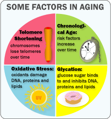
If you are over 60, which I am, your risk dying doubles every 8 years. Research shows that shortening of telomeres only accounts for 4% of the difference. Chronological age and gender account for an additional 33%. The remaining 63% can be attributed to:
Oxidation sounds like a strange contributor to aging. After all, we need oxygen to breathe and a byproduct of this basic life function is oxidants. Oxidants result from oxygen combining with sugar to produce energy and byproducts called free radicals. Not all free radicals are internally produced. They can also come from infections, inflammation, alcohol, smoking, excessive sun exposure, radiation from x-rays and environmental toxins. Free radicals can have a negative impact on individual cells, proteins and fatty tissue. Free radicals over time can build up in the body and are associated with aging. In a recent study the lifespan of worms was increased by 44% by neutralizing oxidants.
Glycation involves excess glucose binding with our DNA, proteins and fats. Excess glucose begins to interfere with normal body tissue functions. The older we get the more the glucose creates health problems contributing to aging let alone body mass. Research shows that restricting calorie intake and selecting foods low in sugar leads to reduced age-related disease and extended lifespans in mice to monkeys.
Inflammation is the body’s natural response against infections and injuries. It also contributes to tissue injury and ultimately to aging. Chronic or persistent inflammation without significant infection is evidence of an immune system that no longer recognizes host body tissue as its own. As we age autoimmune conditions including chronic inflammation become more prevalent. Chronic inflammation destroys normal cells and contributes to the aging of the cardiovascular and nervous system. Inflammation contributes to age-related neurodegenerative diseases, such as Alzheimer’s and Parkinson’s.
Stress harms DNA and speeds the aging process. A study in 2004 showed that psychological stress shortens telomeres in immune cells. Evidence shows that stress, the endocrine system response and the occurrence of disease define age more than chronological aging. Certain diseases occur when anabolic hormone levels start to decline and catabolic hormones start to increase. The latter, such as Cortisol, can contribute to the breakdown of body tissue. As proof of just how much stress contributes to aging and premature death, interviewers who spoke with centenarians found that they exhibited healthy coping strategies in dealing with illness describing their behaviours as accepting, non worrying and taking life one day at a time.
Immune Response refers to the specialized cells generated by our bodies to fight off disease. Researchers in Israel at the Technion studied the natural decline in the immune system as we age. This inability to fight off diseases when we are older is one of the reasons for the statistics about the risks to humans when they reach 60 and over. By suppressing B-lymphocyte immune cells in aging mice and using a drug commonly used to treat rheumatoid arthritis, the mice were able to manufacture healthy replacements using bone marrow. Clinical trials have begun in human populations suffering from B-cell lymphoma.
What are the prospects for human immortality?
The SENS Foundation is dedicated to re-engineering our bodies to end aging through rejuvenation biotechnology. These biotechnologies cover major research areas including cell loss, tissue atrophy, nuclear and mitochondrial mutations, immunotherapy, and targeted ablation. The goals are:
If we eliminate all of these physical processes inherent in aging and develop appropriate delivery systems for restoration and repair some scientists project that we can live 1,000 years. Will someone born in the 21st century be the first millenarian?
]]>If you think of the most recent disasters that made headlines in 2010 and 11 you can begin to envision how robots can play a part in prolonging life in emergencies. From the earthquakes in Haiti and South Island, New Zealand, to trapped miners in Chile, the tsunami in Japan, and most recently the foundering of the cruise ship, Costa Concordia, engineers get inspired to come up with new ways of supplementing human intervention and rescue efforts using robotic systems. In some of these recent tragedies robotic devices were deployed. But that is not the only use for robots in emergency medicine.
The Robot Will See You Now
Robots are used in emergency medicine in many ways. One is as patient screening tools, marrying robotics with computing science. Hospital emergency rooms represent bottlenecks with patients spending long hours waiting for a doctor or nurse. Shortening the wait times using a triage robot has inspired engineers at Vanderbilt University to develop TriageBot,

A smart kiosk, TriageBot takes a medical history, measures vital signs and detects problems. The robot prioritizes patients based on severity of symptoms and history and can connect to on site staff even outside the emergency department should they be needed. The form of TriageBot is still under development. It could look like the fanciful and friendly robot displayed above or resemble an airport check-in terminal, or it could be built into a hospital waiting room chair. Designed to continuously monitor the patient before being seen by a member of the medical team, TriageBot represents an innovative use of robots in hospital settings.
But not all emergencies happen in the waiting room of hospitals. TriageBot systems when hooked up to a telecommunications network can provide both screening as well as monitoring of patients in remote areas. As the software evolves this type of emergency medical robot should prove invaluable.
The Japanese have a way with robotic systems and not to be outdone, Kyushu University has been experimenting with a robot prototype mobile monitoring system. (Click on the hyperlink in the last sentence to watch the movie. Although the narrative is in Japanese, you quickly get an understanding of how this apparatus works.)
Built for AICHI Expo 2005 this mobile chair features remote control driving done by emergency medical staff off site, vital signs monitoring, emergency first aid and even a defibrillator. The remote operator can talk and see the patient, communicate with bystanders and give them information on how they can help a person having a seizure, fainting or heart attack.
The Robot Will Find You Now
CRASAR is the Center for Robot-Assisted Search and Rescue at Texas A&M University. On its website it lists participation in using rescue robots going back to the collapse of the World Trade Center buildings in 2001. One area of development focuses on using robots as human proxies to provide communication with a trapped person that cannot be reached immediately after a disaster.
Survivor Buddy can work with medical staff to interact with a survivor during the period when help is not yet able to extricate the person. The robot includes a monitor that displays non-verbal human attributes by rotating and moving in a human-like way.
Sandia National Laboratories have been working on robots that can deliver food, water, oxygen and medical supplies to trapped miners underground while encountering environments that could prove lethal to human rescue workers. Designed to withstand flooded areas and explosions from pockets of methane gas the Gemini-Scout Mine Rescue Robot comes equipped with a range of sensors as well as pan-and-tilt, and thermal cameras that elevate to see over obstacles. It travels on flexible treads that climb over obstacles. Using a game controller, Gemini-Scout is designed to work with first responders and can be outfitted for earthquakes, fire and other disaster scenarios.

The Snakebot employs biomimicry to do search and rescue for trapped victims of disaster. An active scope camera that slithers, this robot was used to find trapped people under the wreckage of the 2011 earthquake and tsunami that struck Japan.
But snakebots can even go beyond search and rescue. Carnegie Mellon University roboticist, Howie Choset, has designed a snakelike camera device that with 102 joints is capable of imaging and mapping internal organs such as the heart muscle. Although not a search and rescue application, these types of robot designs demonstrate flexibility with capability to serve multiple biomedical applications.
Nowhere have emergency medical robots been studied more than in the military where they are seen as extraction and evacuation tools that reduce collateral casualties. The United States military increasingly uses robots in all of its field operations including drone aircraft as surveillance and strategic strike weapons, as well as robotic land vehicles. With the goal of having 1/3 of its land vehicle fleet unmanned by 2015, robotic medical evacuation will become a common feature. The American army is testing both robotic evacuation and extraction vehicle technology to move patients from fire zones to hospitals. This includes using humanoid robot designs like the one pictured below.

Using robots capable of picking up an injured soldier on a battlefield represents a way of reducing deaths associated with casualty recovery operations.
The military is also testing unmanned aerial systems that can land and working with casualty extraction robots, remove wounded soldiers from environments where radiation or toxic gas would make it impossible for human intervention. Unmanned aircraft are also being tested to provide surveillance and medical response. To provide the human touch, robot extraction systems include telepresence so that a soldier remains connected to a medical support person even though they are not physically on site.
In our next blog we look at the emerging field of robot exoskeleton technology and its biomedical uses.
]]>So where are robots and robotic devices making an impact on biomedicine in the second decade of the 21st century? And where are we going with this type of technology as the century unfolds?
We have already described implantable sensors and devices as well as intelligent prosthetics in a previous blog. In this discussion divided into 5 sections we will look at the following:
Surgical and Diagnostic Robotic Systems
In the last 20 years we have seen the evolution of remote presence technology giving doctors the ability to provide medical help to patients in remote areas of the world. These technologies have proven useful for creating virtual presence for interviewing, patient screening and diagnosis, and medical consultation. One such device is called RP-7, a robot that looks like it came right out of a sci-fi. RP stands for remote presence and gives physicians the ability to see and speak with patients and hospital staff. Through its interface the RP-7 gives a doctor the ability to use electronic stethoscopes, otoscopes and ultrasound and receive transmission of all medical data.

But diagnostic robotic tools like RP-7 are just the beginning. A number of robotic designers have built or are building surgical robots with dexterity that is equal to if not better than the best surgeon. These systems feature 3 or 4 arms, stereoscopic 3D imaging and dexterity that no human surgeon can match. Why is there such a growing interest in these types of devices? Because robotic surgeons increase accuracy, decrease operating times, lead to shorter hospital stays, and produce fewer complications.
Here is a quick history of some of these devices beginning in the 1990s and ending in 2012.
Aesop
Computer Motion, out of Santa Barbara, California, was the creator of Aesop a surgical-assist robot built in the 1990s. The acronym Aesop does not have Greek-authored roots but rather stands for Automated Endoscopic System for Optimal Positioning. Developed to assist surgeons for endoscopies, NASA hoped to use Aesop in space where its better than human dexterity made it highly useful for doing repairs. Aesop was voice-command controlled, much preferred over eye or head-tracking motion control for operating room procedures.
Zeus
This robotic device was a second generation successor to Aesop and was first demonstrated in laboratory studies in 1995. It allowed surgeons to do minimally invasive complex procedures through incisions smaller than the diameter of a pencil. The company that created Zeus eventually merged with the creator of the next system featured in this blog.
da Vinci
Intuitive Systems of Sunnyvale, California, after its merger with Computer Motion, developed a robotic-assisted minimally invasive surgical system called da Vinci(R). Pictured below this robot first received FDA approval in 2000. It features a surgical console with 3D high-resolution magnified viewing connected to 4 interactive highly flexible robotic arms that can be manipulated to perform operations.

Each robotic arm features EndoWrist instruments with a range of motion far greater than a human hand. For many surgeons hand tremors can be a career ending problem but with da Vinci tremors are dramatically lessened with the robotic arms providing enhanced dexterity.
Amadeus
Titan Medical, out of Toronto, Canada, is in the process of developing Amadeus(R), a robotic surgical system designed for remote access surgical procedures. Amadeus gives surgeons the ability to do procedures from anywhere, whether in the operating room or on the other side of the globe. Amadeus lets the surgeon feel the patient through touch feedback technology. Several models are under development with one specifically designed to perform surgery in confined spaces where a surgeon would find it impossible to access the patient.
The Near Future for Surgical Robots
Surgeons have two hands. When more are needed two or more surgeons get involved in an operation. Medical robots are not so limited and more arms means more tools can be brought to bear during a procedure with fewer surgeons needed to operate them. Robotic arms can feature touch feedback systems that provide tactile and pressure sensation to the surgical operator. Robotic arms can be generalists or specialists. One can control a 3D camera that can maneuver in the surgical site providing better than human vision while others can use multiple attached instruments to help a surgeon complete a procedure faster minimizing trauma to the patient. Robotic arms and fingers can be smaller than any human’s arms and fingers. Robotic surgeons can be autonomous entering a body, guided remotely to a surgical site, doing the repair and then exiting.
Are we there yet? Are we near the time when robots will do procedures autonomously without the presence of a surgeon? Not yet but ultimately that is where the technology is headed in the 21st century.
]]>