Today, we have tools to cut out faulty genes and replace them with healthy ones. We can 3D-print human tissue. Does this mean we will soon be able to direct cells to build almost any human organ or part to then integrate into our bodies? What’s holding us back from a Frankensteinian future?
We may face two different outcomes:
It is no longer just imagination. The neuroscience researchers at the University of Wisconsin-Madison are 3D printing functional human brain tissue describing their work as opening a new window into tackling neurological and psychiatric disorders and creating re-engineering treatments for Alzheimer’s and Parkinson’s. What’s not to like?
Sy-Chun Zhang is a professor of neuroscience at Wisconsin’s Waisman Center. He describes what 3D-printing human brain tissue could lead to: “This could be a hugely powerful model to help us understand how brain cells and parts of the brain communicate in humans. It could change the way we look at stem cell biology, neuroscience and the pathogenesis of many neurological and psychiatric disorders.”
Zhang and his associates have used a bio-ink containing human pluripotent stem cells to print stacking layers of brain tissue allowing “the neurons to grow into each other and start talking.” The printed brain tissue includes the cerebral cortex and the striatum. The cells from different brain parts when printed start communicating with each other.
Work being done on tissue regeneration at Tufts University is leading us to the potential to regrow severed or damaged limbs. Michael Levin is a biologist and professor at the university studying cell tissue regeneration and bioelectricity. He runs the Levin Lab with research that goes well beyond uncovering the cellular and genetic mechanisms responsible for tissue growth.
Levin is integrating biological electrical signalling into experiments when working with stem cells to regenerate complex tissues. He sees bioelectricity as the next big step in human tissue regeneration. It’s not quite Frankenstein, but it sounds a lot like it.
Levin theorizes that genome editing technology has limitations. He states by understanding the bioelectricity that binds cellular networks we will start untangling the software code of life.
When the Wisconsin researchers printed the brain cell tissue, the neurons and other cells started communicating using electrical signals. They didn’t understand how this happened, nor did they understand the shared messages being sent by the brain cells.
Levin wants to do a deep dive into the nature of bioelectric messaging in living things. He is co-editor of the journal Bioelectricity, where leading-edge research in this field is being published. He believes that we limit our scientific understanding of how biology works by studying only the genome and DNA. By manipulating bioelectric signals, he believes, it will give our bodies the equivalent of what we do when we adjust the thermostat in our homes to change a room temperature. Today in biology to make that type of adjustment we do the equivalent of rewiring the entire house.
Levin along with serial entrepreneur, Jess Mah, have founded Astonishing Labs, a company that plans to do more research in this area to understand bioelectric messages and the role they play in the body. Can they help to regrow worn and broken parts and extend our body’s healthy lifespan?
]]>Researchers, however, are exploring a future where bioprinting will go below the sub-cellular level in constructing living tissue and biopolymers at a nanoscale. States Professor Seung Soo Oh, of Pohang University in South Korea, “The significance of this research lies in proving for the first time the possibility of printing 100% functionally and structurally active biopolymers in ultrafine 3D structures.”
What are biopolymers? They constitute the building blocks of cells. And now, research being done by Professor Oh and others, published recently in Advanced Science, indicates the development of technology to 3D print biopolymers molecule by molecule.
Pohang isn’t the only place where researchers are pushing the technological limits of 3D bioprinting. Back in 2018 scientists at the Lawrence Berkeley National Laboratory and UC Berkeley invented a way to synthesize DNA making DNA printing a future possibility. With the work at Pohang combined with Berkeley, we are a step closer to bioprinting DNA.
For researchers and bioengineers, the ability to 3D-print DNA opens new opportunities that can potentially revolutionize medicine. Imagine a printer that not only can produce living organs and tissues, but also DNA-specific constructs at the sub-cellular level including RNA, proteins, genes, and individual strings of nucleotides.
If unfamiliar with the term nucleotides, these are the fundamental chemical components of nucleic acids. We have named them adenine (A), thymine (T), guanine (G), and cytosine (C). They pair to form chemical bonds with A and G and C combining to form DNA strands. In other words, they are the building blocks of life as we know it here on Earth.
When you start bioprinting nucleotides, personalized medical therapies take on a whole new meaning. It means you can print healthy genes and replace defective ones that cause congenital diseases.
Where CRISPR/Cas9, also developed at UC Berkeley, gives researchers and medical practitioners the ability to cut out individual gene sequences and replace them with healthy ones, now with DNA bioprinting it becomes possible to print an entire chromosome. Imagine what this could mean in cancer treatment and for many other intractable diseases.
The bioprinting dream, however, goes even further. Using the technology will revolutionize agriculture, pharmaceuticals, manufacturing, and even the fuels we make to create energy. Reinforcing this extended capability, Professor Oh states that 3D bioprinting isn’t restricted to just the “bio.” He sees its use with “various materials with diverse optical and electrical properties, including complex materials such as quantum dots and carbon nanotubes.”
Combining the South Korean advancements in nanoscale 3D printing of biopolymers with the work at Berkeley and biomedical engineers may soon see a future where we will print genes, chromosomes, replacement organs and tissue.
This is not Frankenstein’s monster pieced together and made animate, but rather a technological advancement that will allow us to reconstruct our damaged selves from the DNA level and up and even integrate nanomachines and non-biological components.
When Ray Kurzweil first envisioned what he called the Singularity, it involved the merging of human and artificial intelligence, in other words, what Star Trek would call a “mind meld.” I don’t think Kurzweil envisioned integration at the level of DNA.
His prediction of 2045 as the date when humans would become one with our technology is still 22 years in the future. But the evolution of 3D printing of biology with technology capable of adding integrated nanosized circuits puts a new spin on what the Singularity will be and when it might occur.
]]>The medical field has been experimenting with 3D printing for more than two decades and we are now beginning to realize the full benefits. The technology offers inexpensive alternatives to traditional medical interventions. Today, 3D printed prosthetics and implants for dental, spinal, and other disorders are replacing traditional methods of manufacturing these items. It is even being used to do rapid builds of custom surgical tools to meet the specific needs of a single or multiple medical procedure.
The benefits of this ability to do mass customization to produce complex end products mean better patient interventions and healthier long-term outcomes. That’s the beauty of 3D printing. Timely production, quick alterations, customization on the fly, and one-off items. And the cost when compared to manufacturing the traditional way is significantly less meaning savings for hospitals in terms of the need to keep inventory volumes sufficient for anticipated procedures, with the items often exceeding their shelf expiry dates. And it means those cost savings can be passed on to patients making medical interventions more affordable.
3D printing has introduced bioprinting, using living cells as the ink to create tissue and organs for surgical implantation. It means instead of using donated tissues and organs, the patient’s own cells can be the building blocks to treat organ failure and reconstruction. For hospitals, the chronic shortage of transplantable organs will no longer be a problem. Instead, tissue engineering using the patient’s own stem cells combined with growth factors can produce the living ink that 3D printers can use to build a new kidney or other vital organs. Or 3D printers can apply the living ink to a scaffold made of inert materials that become the structure for tissue regeneration whether it be cartilage, bone, muscle, or blood vessels.
How is customization achieved? Whether it’s the use of stereolithography or other means of 3D printing, the technology has the ability to build artificial limbs and implants for clients. Prosthetics can be made within 24 hours for dental, spinal, and even hip implants. Using 3D printing implants and prosthetics solves the persistent problem for orthopedics of having an insufficient supply of generalized products for procedures. Instead, the printer offers complete customization of design that is a perfect fit for the client.
Creating an accurate anatomical model of a patient’s heart in preparation for surgery is now possible using 3D printers that can use medical diagnostic imaging files from MRIs, CAT scans, and other radiology tools as digital sources. It gives surgeons and medical assistants a better gauge of the biological and physical makeup of the targeted organ allows for better decision-making prior to and during the performance of the procedure.
In the past 2D images from catheterization, fluoroscopy, x-rays and other types of scans formed the basis for a surgeon’s understanding of a patient’s medical status. The alternative to this was the study of the anatomy of cadavers. But finding a cadaver with the same pathology as that of the patient was next to impossible. 3D-rendered anatomical models, however, render those past methodologies obsolescent.
3D printing is fast. The prosthetics and implants produced this way exhibit accurate resolution and a level of accuracy not possible from milling and forging an alternative. And delivery time for the finished object is a fraction of what it would be if a manufacturer were to be asked to provide an equivalent.
Finally, it should be noted that medical 3D printing brings democratization of design to medical interventions as well as an unprecedented ability for knowledge sharing among medical professionals and researchers.
The use of 3D-printing data files means what is produced can be shared allowing for faster dissemination of new knowledge and techniques within medicine. Medical practitioners and laboratory researchers can access downloadable files from open-source databases to replicate 3D-printed bio-inks, models, and devices. Since 2014, the United States National Institutes of Health have established a 3D Print Exchange to promote sharing of 3D-print files in support of biomedical science advances.
]]>| July 7, 2020 – In the latest missive from Peter Diamandis, he focuses on advances in our medical and scientific knowledge around stem cells, organ generation, bioprinting, and extending human lifespan. Peter has picked an interesting time to be full of optimism about the future of our species as we face a current pandemic and the mounting threat of anthropogenic atmospheric and ocean warming. He justifies this optimism because he sees disruptive forces of innovation breaking the back of our current problems, and is looking to a far brighter future for the planet and us. Here are mostly his words and ideas which I have edited where warranted to share with my readers. I hope you find the subject intriguing and maybe even a bit disturbing. As always your comments are appreciated.
Lizards can regrow entire limbs. Flatworms, starfish, and sea cucumbers can regrow entire bodies. Sharks replace lost teeth, as many as 20,000 throughout a lifespan. So why can’t we do the same? The answer: Through cutting-edge innovations in regenerative medicine, research is underway to do just that. In 2019, investors pumped $10 billion into this field, and currently, there are over 1,000 clinical trials underway trying regenerative treatments on 60,000 patients. As Big Data and Artificial Intelligence (AI) transform medicine, regenerative medicine will take us to these three Rs:
1. ReplenishStem cells are undifferentiated cells that transform into specialized cells from embryos to when we are born and beyond. They can be found in many areas of the body. Stem cells differentiate to become heart, neurons, liver, lung, skin and other specialized tissues. They also can divide and produce more stem cells. As a child or young adult, we have stem cells in large supply. They trigger when the body needs to repair itself. They are summoned to sites where there is damage or inflammation. As we age, the supply of stem cells diminishes by as much as 100- to 10,000-fold throughout different tissues and organs. Stem cells also undergo genetic mutations over a lifespan which reduces their quality and effectiveness at repairing damage. If you could restore and rejuvenate a body’s stem cell population we can extend our healthy years dramatically. One way to do this would be to extract and concentrate our autologous adult stem cells harvested from adipose (fat) tissue or bone marrow. Another source comes from umbilical cords and placentas kept after birth. The advantage of the latter over the former is these stem cells have not been subject to aging like those found in adult adipose tissue or bone marrow. Umbilical and placenta stem cells provide the undamaged software of a newborn and when cultured to multiply, can be injected into joints or administered intravenously as a rejuvenation therapy. Using these stem cells can help to fight inflammation, autoimmune disease, increase muscle mass, repair joints, and even revitalize skin and grow hair. A burgeoning field, stem cell research has seen a 40-fold increase in scientific publications over the last few years. The market globally is estimated to grow to $14.8 billion USD by 2022, growing at an annual growth rate of 14.7%. Examples of regenerative treatments include:
2. ReplaceEvery 10 minutes an American is added to an organ transplant waiting list. As of March 2020, 112,000 people were waiting for replacement organs. Countless more people never make it to the waitlist with an average of 20 dying each day. The result, 35% or approximately 900,000 people can be prevented from dying with better and more timely access to replacement organs. Promising a remedy to organ shortages, replacement and regenerative medicine represents a massive opportunity. The opportunity has led to emerging organ entrepreneurs. One of these is United Therapeutics, started by Dr. Martine Rothblatt, a one-time aerospace entrepreneur and founder of Sirius Satellite Radio. Personal circumstances led to a change of careers when Rothblatt’s daughter developed a rare lung disease. Her “Moonshot” project to replace her daughter’s unhealthy lungs, and others with similar affliction, has become a major business opportunity pursuing three disruptive strategies that could change medical transplants forever. Rothblatt’s company is United Therapeutics where she is CEO. With an initial focus on diseases of the lung, she has set out to create replacement lungs. To accomplish this goal, she has pursued a number of technologies in parallel including. A. 3D Printed OrgansIn 2017 she teamed up with one of the world’s largest 3D printing companies, 3D Systems, to build a collagen bioprinter. She teamed up with another company, 3Scan, to create maps of the lungs structures and interior using detailed micro slices programmed into a bioprinter. An ultraviolet laser passing light through a shallow pool of collagen doped with photosensitive molecules served as the medium for the build. The laser targeted the areas identified by the mapping causing collagen to cure and form a scaffold to which human cells attach. So far the technology has been able to produce lung structures with resolutions of 20 micrometers. The end goal is to get to one micrometer to achieve a functioning replacement for a human lung. Once a scaffold capable of these dimensions can be built it will be infused with living stem cells using recellularization. Stem cells will grow and differentiate, populating the scaffolding, and producing a functional lung. Can it be done? In 2018, Harald Ott, an experimental surgeon at Harvard University reported that he had pumped billions of human cells (from umbilical cords and diced lungs) into a pig lung stripped of its own cells. When Ott’s team reconnected it to a pig’s circulation, the resulting organ displayed rudimentary lung function. B. XenotransplantationRothblatt is pursuing the transplanting of animal organs into humans which is known as xenotransplantation. Because an adult pig has organs similar in size and shape to those of humans, her company is focusing on genetically engineering pigs to harvest their organs for human transplants. “It’s actually not rocket science,” said Rothblatt in a 2015 TED talk. “It’s editing one gene after another.” To accomplish this goal, her company has made investments in Revivicor Inc. and Synthetic Genomics Inc. signing funding agreements with the Universities of Maryland, and Alabama, and New York-Presbyterian/Columbia University Medical Center to start a xenotransplantation program for hearts, kidneys and lungs. Rothblatt hopes to see the final results in three to four years. The end goal for the company will be to produce up to 1,000 sets of healthy pig lungs, called xenolungs, annually from genetically engineered pigs. C. Ex Vivo PerfusionToday only 30% of human-donated lungs meet transplant criteria. Of these, 85% remain usable upon arrival at a surgical site for implantation. The end result, nearly 75% of donated lung tissues never get used by recipients in need of a transplant. But what if the donated lung tissue could be rejuvenated? In 2016, Rothblatt invested $41.8 million in TransMedics Inc., an Andover, Massachusetts company that has developed the XVIVO Perfusion System which takes marginal-quality lungs initially fail to meet transplantation standard-of-care criteria and perfuses and ventilates them in normothermic conditions, to provide an opportunity for surgeons to reassess their transplant suitability. The perfusion system can also be used with heart, kidney, and liver tissue for transplantation. RejuvenateResearchers at Stanford and Harvard University have demonstrated that older animals when transfused with blood from young ones experience regeneration in many of their tissues and organs. The opposite is true as well that when young animals are transfused with blood from older donors, they experience accelerated aging. Elevian (a Harvard University spinoff) is using this “young blood” effect in its approach to studying longevity. CEO Mark Allen, MD, is among the founders along with a dozen MDs and PhDs from the university. They have identified specific factors responsible for the effect described in the previous paragraph. A naturally occurring molecule known as “growth differentiation factor 11,” or GDF11, which, when injected into older mice, reproduces regenerative effects in the heart, brain, muscles, lungs, and kidneys. Further, GDF11 reduces age-related cardiac hypertrophy, accelerates skeletal muscle repair, boosts exercise capacity, increases brain function and cerebral blood flow, and even improves metabolism. From this Elevian is developing a number of therapeutics to regulate GDF11 and other circulating factors with a goal to restore the body’s natural regenerative capacity. The company believes with its current research it can counter the root causes of age-associated disease promising to reverse or prevent many of them and to extend a healthy lifespan. Final ThoughtsIn 1992, the futurist Leland Kaiser created the term regenerative medicine. He called it “A new branch of medicine…that attempts to change the course of chronic disease and in many instances will regenerate tired and failing organ systems.” From these initial roots has grown an industry that in 2018 generated $23.8 billion USD and is projected to exceed $151 billion by 2026. If the work described here pans out, the road ahead for improvements to human health will be enormous. Imagine having the ability to regenerate, replenish, and replace entire organs and metabolic systems on command. Are there unintended consequences to Peter’s hopeful description of the biomedical breakthroughs that lie ahead? Extending the natural lifespan and making for a longer and healthier lives in a person’s 70s, 80s, 90s, and even 100s and beyond comes with an added burden to the planet which already has 7.8 billion people on it. What are the implications for work and retirement? What are the implications for subsequent generations seeking the jobs of those who are older and no longer prepared to retire because of aging and disease? At the beginning of the 20th century, the average human lifespan was 40. By the beginning of the 21st century, it had almost doubled. By 2100, will it double again? Peter Diamandis is convinced it will and we are seeing in this article the beginnings of the technologies that will make that a reality. |
This invention represents a rapid departure from the current standard of care for patients with severe large-area burns. The standard practice involves autologous skin grafts or acellular substitutes and is fraught with challenges, particularly susceptibility to infection. And any existing bioprinters are not portable, but rather are bulky, expensive and slow. That’s not the case for this new handheld instrument that delivers mesenchymal stem cells contained in fibrin sheets directly to a wound surface along with bio-neutral polymers and proteins to evenly distribute cell layers. The end result, improved surface skin, and epidermal regeneration. A paper describing the invention appears in the February 4, 2020 edition of the journal Biofabrication.
Burns are a global public health problem, according to the World Health Organization, accounting for an estimated 180,000 deaths annually with most occurring in poorer countries. And while burn death rates are declining in rich countries, the rate of child deaths from burns in the Developing World is seven times higher than in the Developed World. In 2004, almost 11 million people globally suffered from burns severe enough to require medical attention.
The inspiration behind the invention of the portable bioprinter was seen by the University of Toronto and Sunnybrook team, to be the need to address a lack of expertise and facilities to deal with burns in the Developing World.
Where surgically transplanted skin grafts require a significant amount of healthy donor skin to be applied in deep wounds to all three layers of the skin, the epidermis, dermis, and hypodermis, availability is often not there.
But the handheld bioprinter with minimal training can deposit epidermal and dermal tissue along with a mixture of bio-neutral polymers and proteins for even distribution of the cells layers. The printer also delivers hyaluronate, a protein that is contained in hyaluronic acid to promote healing and skin cell integrity. The combination contained within the bioink in mouse and pig studies showed effective repair results of burn wounds. It soon may be available for human trials and become a new standard in dealing with deep-wound burns, potentially replacing skin grafting processes.

In a Scientific Reports paper published on February 12th of this year entitled “In Situ Bioprinting of Autologous Skin Cells Accelerates Wound Healing of Extensive Excisional Full-Thickness Wounds,” the authors at the Wake Forest School of Medicine describe how over 7 million patients in the United States experience chronic and episodic wounds leading to $25 billion in annual expenses. Chronic wounds include diabetic, venous, and pressure ulcers. Episodic wounds come from burns and other external non-disease sources. They note that “full thickness” skin injuries are a major source of morbidity and mortality. More than 500,000 Americans annually suffer from burns requiring in-hospital treatment including skin grafts, and for the U.S. military, between 10 and 30% of combat casualties are a result of burn injuries.
The earlier skin wounds get treated leads to greater survivability from burn-related injuries. Currently, skin grafts are the gold standard but getting healthy donor grafts is fraught with potential challenges both in availability and in terms of immune rejection risks. Synthetic materials represent a substitute for grafts but they are costly and cosmetically produce less than desirable results.
A number of other types of technologies have been attempted to heal wounds and accelerate healing. These include spraying or applying liquids with skin cell tissue on wound sites to seed the healing process. The bioprinter takes the above processes several steps further.

Today’s 3D bioprinters are limited by several constraints. The first involves the bioink. The second involves the print heads that inject the bioink. The third involves the medium in which to lay down the ink. The bioink has to nourish the living cells contained within the solution. The print heads must be capable of delivering the bioink solutions keeping the living cells viable. And finally, the medium in which the bioink is deposited must be such that it forms a structural matrix for the growth of the cells to meet the required organ or blood vessel being constructed.
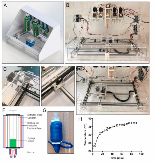
The U. of T. paper describes a functional 3D printer built from custom-designed parts combined with open source controller boards and motors with five print heads, and a Cartesian coordinate XYZ system to plot the 3-dimensional print job where X is width, Y is depth, and Z is height. Traditional inkjet printers are designed for 2D output laying down ink on a sheet of paper. But the added Z dimension is the one that presents the biggest challenge because the method of printing with bioink lays down material using the X and Y axis and then systematically adds layer upon layer to create the Z. Another feature of this open source printer is the print bed which allows the operators to switch out different printing surfaces to address issues like fine temperature regulation during organ or blood vessel construction.
The bioink contains gelatin and readily available Pluronic F-127, a bioreagent suitable for culturing living cells. When doing bioprinting it involves two stages. First, you lay down a material that forms the matrix design for the organ and blood vessel to be constructed. Then second, you follow that with the bioink. The first material is not meant to be permanent. Once the desired organ or blood vessel is constructed, a third step involves flushing away the material in the first step and leaving finished living tissue.
A relative bargain, the cost of the printer amounted to $3,000 CDN in parts and materials. Resolution of the print is between 0.5 and 1 millimeter. In testing the printer was capable of replicating complex vascular tissues with consistency and accuracy.